
Fall 2019 Schedule
Instructor: Dr. C. Hays, Office: Science 2032
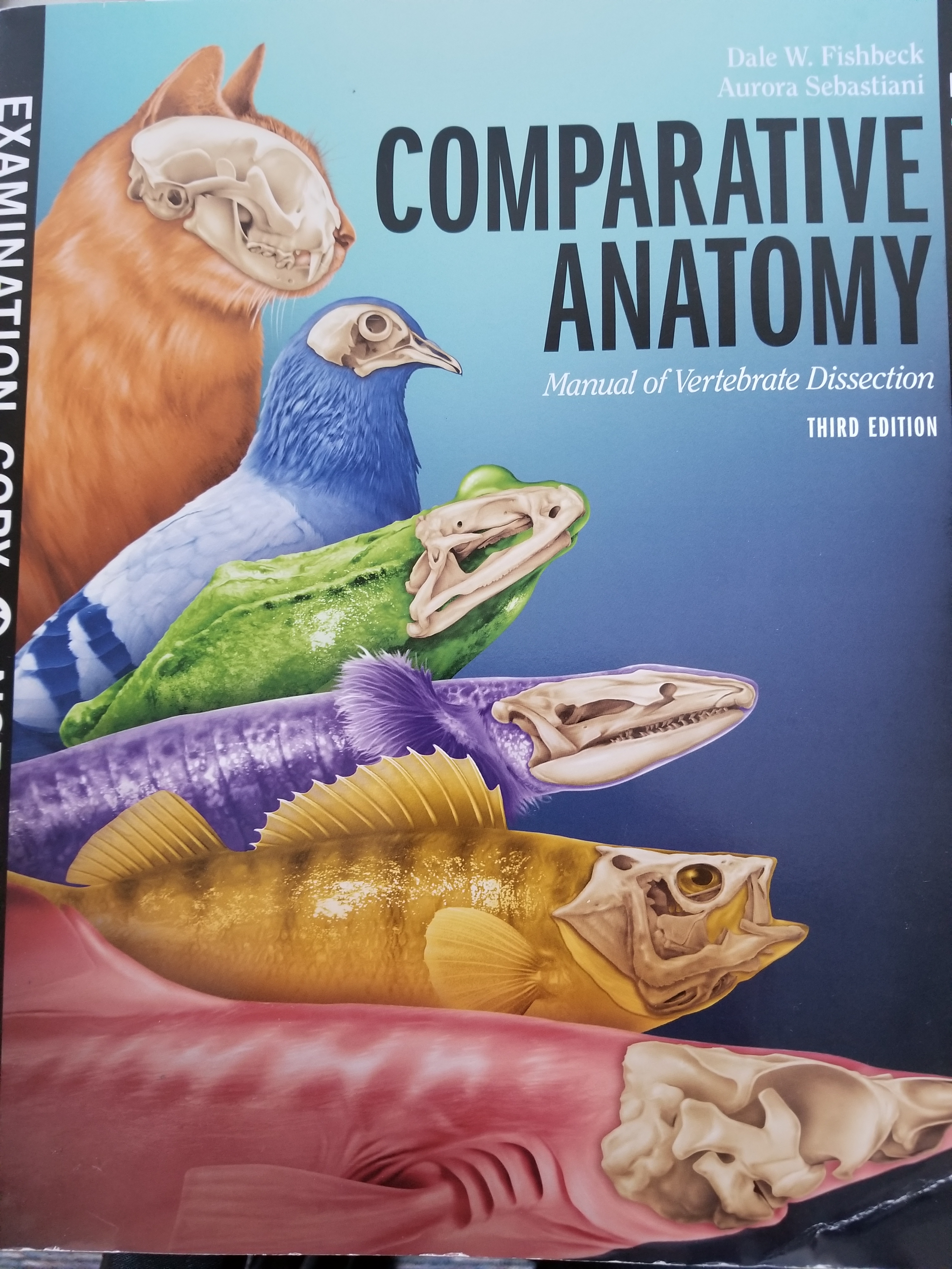
REQUIRED: Comparative Anatomy Manual, of Vertebrate Dissection, 3rd Edition, Fishbeck & Sebastiani, Morton Publishing Company, 2015. ISBN 978-1-61731-042-3
Dissecting Kit. Available in bookstore; includes a scalpel with replaceable blades, a blunt probe, and small scissors.
Not required, but strongly recommended, are disposable gloves and a lab coat or an old shirt to protect your clothing. Respirators with filters and eye goggles are available upon request.
Note the link at the bottom of this page for all answers to the laboratory objectives.
THE PROCHORDATE, THE LAMPREY, INTEGUMENT
AUGUST 17,19 – THE PROCHORDATE, THE LAMPREY, INTEGUMENT
CHAPTER OBJECTIVES:
Introduction
1. List five fundamental characteristics of a chordate.
2. Know the following directional terms and planes: dorsal, ventral, caudal, cranial, anterior, posterior, medial, lateral, proximal, distal, sagittal, mid-sagittal, transverse and frontal.
Chapter 3
1. Classify Amphioxus (=Branchiostoma) in the appropriate phylum and subphylum.
2. Why do we study Amphioxus if it is not a vertebrate?
3. Define myomere.
4. Where does the mature Amphioxus spend most of their “time”?
Chapter 14
1. Classify lampreys in the appropriate phylum and subphylum.
2. Describe the vertebrates that belong in this group.
3. What is the name of the larval form of the lamprey?
4. Briefly, describe the life cycle of a lamprey.
5. Describe the jaw of lamprey. Define agnathan.
6. What is the difference between the terms myotome and
myomere?
Chapter 4 & & 15 & 24 & 52
1. Are fish scales embedded in the epidermis or dermis ?
2. Name the two skin layers.
3. Characterize keratin.
4. List some examples of epidermal derivatives in mammals.
Introduction: Read this chapter and review anatomical terminology on p.5.
View a Nova film (50 minutes) during your lab period.
Chapter 3: Read this chapter and Observe a whole mount of Amphioxus. Identify the following:
Dorsal fin, Myomeres, Myosepta (myoseptum sing.), Notochord, Nerve cord, Oral hood, Pharynx with Pharyngeal slits, Midgut, Hindgut, Anus.
Chapter 14: Read this chapter and observe a whole mount of a Lamprey. Identify the following:
Nostril, Mouth (Buccal funnel), External gill slits, Myomeres, Myosepta, Heart, Dorsal Aorta, Notochord, Spinal cord, Brain.
Observe a whole mount of Ammocoetes larva. Identify the following:
Oral hood (with tentacles or lobes), Velum, Gill filaments (lamellae), Gill pouch, External gill slits, Intestine, Anus, Liver, Gall bladder, Heart, Brain, Spinal cord, Eye, Notochord, Myomeres (actually myotomes), Dorsal fin, Caudal fin.
Observe a cross section through the pharynx of Ammocoetes. Identify the following:
Pharynx, Gill lamellae or filaments, External gill slit, Notochord, Spinal cord.
Chapter 4: Read this chapter and observe slides of: Placoid, Ganoid, Ctenoid and Cycloid scales. Also see figures 4.2, 15.11, 15.12, 15.13, 24.6.
Observe slide of mammalian skin and identify: Epidermis, Dermis, Hair root, Hair shaft. See figures 52.2, 52.3, 52.4.
THE SKELETON
AUGUST 24,26, 31,SEPTEMBER 2,9,14,16 – THE SKELETON
{Suggested timeline: 3 labs for skull, 1 lab for trunk (vertebrae, ribs, sternum), 3 labs for girdles and limbs.}
CHAPTER OBJECTIVES:
Chapter 5, 16, 24, 26, 35, 44, 53
1. Generally, list some functions of the skeletal system.
2. Describe the difference between the axial and appendicular divisions of the skeletal system.
3. Define girdle.
4. Contrast the difference in the embryonic origin of dermal and endochondral bone.
5. Classify the chondrocranium, splanchnocranium and dermatocranium as dermal or endochondral bone.
6. Define fissure, foramen and foramina.
7. Define polyphyodont, diphyodont.
8. What is the dental formula of the cat?
9. Differentiate a primary palate from a secondary palate.
10. Define turbinates and conchae. Describe their functions.
11. Define suture as it relates to the skull.
12. Describe a simple method of telling caudal vertebrae from the other types of vertebrae.
13. Identify two phylogenetic trends in the vertebral column.
14. Describe the advantage of having cervical vertebra(e).
15. What is the function of the sacral vertebra(e)?
16. What is meant by a bicipital rib? Name the two articulations.
17. What is a urostyle? A pygostyle?
18. Why is the bird vertebral column a good example of form matching function?
19. What bones comprise the synsacrum?
20. What is the term used for fin supportive elements?
21. Would one find a clasper on male or female sharks? What is its function?
22. What is the singular form of “phalanges?”
23. What is the innominate bone? Describe the position of the three bones that form the os coxae in tetrapods.
24. Describe some overall patterns of evolutionary change of the skeleton.
SKULL
Observe the following structures on Squalus head skeleton (Chapter 16):
CHONDROCRANIUM (Neurocranium):
Rostrum, Nasal capsule, Eye orbits, Preorbital process, Supraorbital crest, Postorbital process, Otic (auditory) capsule, Precerebral cavity, Rostral fenestrae, Epiphyseal foramen, Superficial ophthalmic foramina, Foramen magnum, Occipital condyles, Vagus foramen, Glossopharyngeal foramen, Basitrabecular process, Basal plate, Optic foramen, Trigeminofacial foramen.
SPLANCHNOCRANIUM:
Seven visceral arches, Branchial arches, Pharyngobranchial, Epibranchial, Ceratobranchial, Hypobranchial, Basibranchial, Gill rays, Gill rakers, Mandibular arch, Palatoquadrate cartilage, Meckel’s = Mandibular Cartilage, Hyoid arch, Hyomandibular cartilage, Ceratohyal, Basihyal.
Observe the following on an Amia skull (or other bony fish) (Use handout provided and Chapter 24):
DERMATOCRANIUM:
Orbit, Maxilla, Opercular, Dentary. These are the same in Amia as in the Perch on page 203.
SPLANCHNOCRANIUM:
Quadrate, Hyomandibular. Use the Amia handout for these.
Observe the following on a Necturus skull (Chapter 26):
NEUROCRANIUM and DERMATOCRANIUM and SPLANCHNOCRANIUM:
Ethmoid plate, Quadrate, Exoccipital, Occipital condyle, Foramen magnum, Premaxilla, Frontal, Parietal, Orbit, Squamosal, Parasphenoid, Vomer, Pterygoid, Dentary, Angular, Splenial.
Observe the following on a Chelydra skull (Use handout provided):
DERMATOCRANIUM:
External nares, Orbits, Premaxilla, Maxilla, Zygomatic (Jugal), Ear cavity, Frontal, Parietal, Squamosal, Temporal fenestrae, Pterygoid, Palatine, Vomer, Internal nares, Surangular, Dentary, Angular, Prearticular, Coronoid, Splenial.
NEUROCRANIUM:
Foramen magnum, Supraoccipital, Occipital condyle, Basioccipital, Exoccipital, Basisphenoid.
SPLANCHNOCRANIUM:
Quadrate, Articular.
Observe the following on a cat skull (Chapter 53):
DERMATOCRANIUM and NEUROCRANIUM:
Premaxilla, Maxilla, Palatine process of the maxilla, Palatine, Lacrimal, Ethmoid, Vomer, Frontal, Orbit, Zygomatic (= Malar =Jugal), Zygomatic arch, Parietal, Occipital, Basioccipital, Occipital condyle, Foramen magnum, Basisphenoid, Alisphenoid, Pterygoid process, Presphenoid, Temporal, Tympanic ( = Auditory) bulla, External auditory meatus, Nasal cavity with turbinates (=conchae), Nasal, Optic foramen, Orbital fissure, Foramen rotundum, Foramen ovale, Sphenopalatine foramen, Anterior palatine foramen, Infraorbital foramen, Jugular foramen, Nasolacrimal canal, Mandible, Dentary bones, Condyloid process, Mandibular body, Mandibular ramus, Mental foramina, Coronoid process, Angular process, Mandibular foramen, Incisors, Canine tooth, Premolars, Molar.
SPLANCHNOCRANIUM:
Observe the hyoid bone of the cat.
TRUNK
Observe a cross section of a Squalus tail (Chapter 16). Identify the Nerve cord in the Neural or Vertebral arch, Notochord remnant in centrum, Caudal Blood Vessels in the Hemal arch. See Figure 16.13
Observe mounted fish skeletons and identify the following (Chapter 24):
Trunk vertebrae, Caudal vertebrae, Vertebral body = Centrum, Hemal arch & Spine, Neural arch & Spine, Ribs.
Observe Necturus skeletons and identify (Chapter 26):
Cervical vertebra = Atlas, Trunk vertebrae, Sacral vertebra, Caudal vertebrae, Hemal arches, Transverse processes, Vertebral =Neural arch, Vertebral canal, Neural spine, Cranial zygapophyses = Prezygapophyses, Caudal zygapophyses = Postzygapophyses, Vertebral body = Centrum, Ribs.
Identify the Urostyle on frog skeleton (Figure 35.1).
Identify the following on turtle dorsal shell: Carapace, Vertebrae, Ribs.
Identify the following on bird skeletons (Chapter 44): Cervical vertebrae, Fused thoracic vertebrae, Vertebral section of ribs = Costal ribs, Uncinate processes, Sternal section of ribs = Sternal ribs, Synsacrum, Free caudal vertebrae, Pygostyle.
Identify the following on the cat (Chapter 53):
Cervical vertebrae, Thoracic vertebrae, Lumbar vertebrae, Sacral vertebrae, Caudal vertebrae, Intervertebral discs, Vertebral body = Centrum, Neural canal = Vertebral canal, Lamina, Spinous process, Pedicle, Transverse Process (=Diapophysis in thoracic region), Cranial zygapophyses = Prezygapophyses, Caudal zygapophyses = Postzygapophyses, Atlas, Transverse foramen, Axis, Dens = Odontoid process, Pleurapophysis, Mammillary process, Sacrum, Sternum (8 sternebrae), Rib: Head = Capitulum, Tuberculum, Neck, Body,
APPENDICULAR SKELETON – GIRDLES and LIMBS
Identify the following on Squalus specimens (Chapter 16):
Pectoral Girdle & Fin: Coracoid bar, Scapular cartilage, Glenoid surface, Suprascapular cartilage, Pterygiophores [Basal & Radial], Ceratotrichia-Cartilaginous fish or Lepidotrichia-Bony fish.
Pelvic Girdle & Fin: Puboischiadic bar, Basal pterygiophores, Radial pterygiophores, Ceratotrichia-Cartilaginous fish or Lepidotrichia-Bony fish, Clasper.
Identify the following on Necturus skeleton(Chapter 26):
Pectoral Girdle & Appendage: Scapula, Humerus, Radius, Ulna, Carpals, Metacarpals, Phalanges.
Pelvic Girdle & Appendage: Ilium, Pubis, Ischium, Acetabulum, Femur, Tibia, Fibula, Tarsals, Metatarsals, Phalanges.
Observe the following on bird skeletons (Chapter 44):
Pectoral Girdle and Appendage: Scapula, Procoracoid = Anterior coracoid, Clavicle, Interclavicle, Sternum, Keel = Carina, Humerus, Ulna, Radius, Carpometacarpus, Phalanges, Digits I, II, III. (note, the most proximal is I and the longest is II)
Pelvic Girdle and Appendage: Ilium, Ischium, Pubis, Acetabulum, Femur, Patella, Tibiotarsus, Fibula, Tarsometatarsus, Phalanges, Digits I, II, III, IV.
Identify the following on cat skeletons (Chapter 53):
Pectoral Girdle and Appendage: Scapula: Glenoid fossa, Coracoid process, Scapular spine, Infraspinous fossa, Supraspinous fossa, Acromion process, Metacromion, Subscapular fossa. Clavicle. Humerus: Head, Lesser tuberosity (= tubercle), Greater tuberosity (= tubercle), Bicipital groove, Condyles -> Trochlea and Capitulum, Medial and lateral epicondyles, Supracondyloid foramen, Olecranon fossa. Radius: Head, Radial = Bicipital tuberosity, Styloid process. Ulna: Olecranon, Semilunar notch, Coronoid process, Radial notch, Styloid process. Carpal bones. Metacarpals. Phalanges: Claws.
Pelvic Girdle and Appendage: Os coxae = Innominate bones: Ilium, Crest of ilium, Ischium, Tuberosity of ischium, Pubis, Acetabulum, Obturator foramen. Femur: Head, Greater trochanter, Lesser trochanter, Trochanteric fossa, Linea aspera, Lateral and Medial condyles, Intercondyloid fossa, Lateral and Medial epicondyles. Patella. Tibia: Condyles, Tibial Tuberosity, Tibial crest, Medial malleolus. Fibula: Head, Lateral malleolus. Tarsal bones: Talus,Calcaneus. Metatarsals. Phalanges: Claw.
SEPTEMBER 21- LAB EXAM 1
MUSCLES
SEPTEMBER 23, 28,30, OCTOBER 5,7,12 – THE MUSCULAR SYSTEM
{Suggested timeline: 1 lab for shark muscles, 1 lab for mud puppy muscles, 3 labs for cat muscles, 1 lab for review.}
CHAPTER OBJECTIVES:
Chapter 6, 17, 27, 54:
1. Compare and contrast the three muscle tissue types.
2. Define belly, origin, insertion.
3. Describe the following muscle actions: Flex, Extend, Abduct, Adduct, Protract, Retract, Rotate.
4. Define prime mover, syngergist, fixator and antagonist.
5. Describe the location of hypobranchial muscles in the shark. What does “branch-” mean?
6. Define the roots “coraco-“, “cleido-“, and “genio-“.
7. Which muscles comprise the quadriceps muscles? Which muscles comprise the hamstring muscles? What muscles comprise the Triceps surae?
Dissect Squalus as described in you lab manual and identify the following external structures and muscles (Chapter 17):
AXIAL MUSCLES: Myomeres, Myosepta, Linea alba, Horizontal septum, Epaxial musculature, Hypaxial musculature.
APPENDICULAR MUSCLES: Extensors (=abductors) and Flexors (=adductors) of Pectoral and Pelvic Fins.
BRANCHIOMERIC MUSCLES: Adductor mandibulae, Intermandibularis, Levator hyomandibulae, Levator hyoideus, Interhyoideus, Cucullaris, Dorsal and Ventral superficial branchial constrictors of Visceral Arches III-VII.
HYPOBRANCHIAL MUSCLES: Coracomandibularis, Coracoarcual, Coracohyoid.
Dissect Necturus as described, and identify the following (Chapter 27):
AXIAL MUSCLES: Myomeres, Myosepta, Linea alba, Horizontal septum, Epaxial musculature, Hypaxial musculature, Dorsalis trunci, Rectus abdominis, External oblique, Internal oblique, Transversus abdominis.
HEAD MUSCLES (Hypobranchial & Branchiomeric): Adductor mandibulae anterior, Adductor mandibulae externus, Depressor mandibulae, Branchiohyoideus, Levator arcuum, Intermandibularis, Interhyoideus, Sphincter colli, Geniohyoideus, Rectus cervicis.
APPENDICULAR MUSCLES: Procoracohumeralis, Supracoracoideus, Pectoralis, Pectoriscapularis, Cucullaris, Scapular deltoid (Dorsalis scapulae), Latissimus dorsi, Triceps brachii, Coracobrachialis, Humeroantebrachialis, Forearm extensors, Forearm flexors, Puboischiofemoralis externus, Puboischiotibialis, Puboischiofemoralis internus, Pubotibialis, Iliotibialis, Ilioextensorius, Iliofibularis, Shank (= hind limb) extensors, Shank flexors.
Dissect the cat as described and identify the following (Chapter 54):
Here are a couple of websites with nice photographs of cat muscles:
https://homes.bio.psu.edu/faculty/strauss/anatomy/musc/muscular.htm
CUTANEOUS MUSCLES: Cutaneous maximus, Platysma.
SUPERFICIAL THORACIC MUSCLES: Pectoantebrachialis, Pectoralis major, Pectoralis minor, Xiphihumeralis.
ABDOMINAL MUSCLES: External oblique, Internal oblique, Transversus abdominis, Rectus abdominis, Linea alba.
DEEP THORACIC MUSCLES: Serratus ventralis, External intercostal, Internal intercostal.
SUPERFICIAL BACK MUSCLES: Clavotrapezius, Clavobrachialis ( = Clavodeltoid), Trapezius (Acromio-, Spino-portions), Latissimus dorsi.
NECK MUSCLES: Sternomastoid, Cleidomastoid, Sternohyoid, Sternothyroid, Thyrohyoid, Mylohyoid, Geniohyoid.
DEEP NECK & BACK MUSCLES: Rhomboideus capitis, Rhomboideus cervicis & thoracis, Splenius.
MUSCLES OF THE HEAD: Masseter, Temporalis, Digastric.
SHOULDER MUSCLES: Supraspinatus, Infraspinatus, Teres major, Levator Scapulae Ventralis, Acromiodeltoid, Spinodeltoid, Clavobrachialis ( = Clavodeltoid), Subscapularis.
PECTORAL LIMB MUSCLES: Epitrochlearis, Biceps brachii, Triceps brachii (Lateral, Long, Medial Heads), Brachialis, Brachioradialis, Extensor carpi radialis longus, Extensor carpi radialis brevis, Extensor digitorum communis, Extensor digitorum lateralis, Extensor carpi ulnaris, Flexor carpi ulnaris, Flexor digitorum superficialis ( = Palmaris longus), Flexor carpi radialis, Pronator teres, Flexor digitorum profundus (5 heads, don’t know the heads by name).
THIGH MUSCLES: Sartorius, Gracilis, Biceps femoris, Caudofemoralis, Semitendinosus, Semimembranosus, Adductor femoris, Adductor longus, Pectineus, Tensor fasciae latae, Quadriceps femoris — Vastus lateralis, Rectus femoris, Vastus medialis, Vastus intermedius.
SHANK MUSCLES: Tibialis cranialis (= anterior), Extensor digitorum longus, Peroneus group, Flexor digitorum longus, Flexor hallucis longus, Tibialis caudalis ( = posterior), Gastrocnemius, Plantaris, Soleus, Achilles tendon.
HIP MUSCLES: Gluteus maximus, Gluteus medius.
DIGESTIVE & RESPIRATORY SYSTEMS
OCTOBER 14,19 – DIGESTIVE AND RESPIRATORY SYSTEMS
CHAPTER OBJECTIVES:
Chapter 8, 18, 28, 55
1. What are mesenteries? What are their functions?
2. Define viscera.
CHAPTER OBJECTIVES:
Chapters 8, 19, 29, 56
1. Describe the overall process of digestion.
2. Name two different structures that aid in mechanical breakdown of food.
3. What is the function of the pharynx?
4. List two methods vertebrates utilize to maximize the efficiency of digestion.
5. List structures of the alimentary canal that can increase surface area.
6. Describe some functions of the liver.
7. Name one reason why shark livers contain so much oil.
8. Explain why the shark’s tongue is not a “true” tongue.
9. Define sphincter.
10. Describe the overall function of the respiratory system.
11. What is a spiracle?
12. Can a vertebrate live on land without lungs? Explain your answer.
13. List the three parts of the mammalian small intestine.
14. Name the three pairs of salivary glands in the cat.
15. What is the function of the shark’s rectal gland.
Chapter 18: Dissect Squalus and observe the following: Parietal peritoneum, Visceral peritoneum, Pleuroperitoneal cavity, Transverse septum, Mesentery proper.
Chapter 19: Dissect Squalus and observe the following: Teeth, Oral cavity, Tongue, Pharynx, Esophagus, Esophageal papillae, Stomach, Gastric rugae, Body of stomach, Cardiac region of stomach, Pyloric region of stomach, Pyloric sphincter, Greater curvature, Lesser curvature, Valvular intestine, Spiral valve, Cloaca, Rectal gland, Liver, Gall bladder, Bile duct, Pancreas, Spleen, Spiracle, Internal gill slit, External gill slit, Gill rakers, Interbranchial septum, Gill filaments ( = lamellae).
Chapter 28: Dissect Necturus and observe the following: Parietal peritoneum, Visceral peritoneum, Pleuroperitoneal cavity, Transverse septum, Mesentery proper, Falciform ligament.
Chapter 29: Dissect Necturus and observe the following: Oral cavity, Teeth, Tongue, Pharynx, Esophagus, Stomach, Pyloric sphincter, Gastric rugae, Small intestine, Large intestine, Cloaca, Liver, Gall bladder, Pancreas, Spleen, External gills, Gill slits, Lung.
Chapter 55: Dissect the cat and observe the following: Diaphragm, Pleural cavity, Parietal pleura, Visceral pleura, Mediastinum, Peritoneal cavity, Parietal peritoneum, Visceral peritoneum, Falciform ligament, Greater omentum, Lesser omentum, Mesentery.
Chapter 56: Dissect the cat and observe the following: Parotid salivary gland, Parotid duct, Mandibular salivary gland, Sublingual salivary gland, Oral cavity, Tongue, Hard palate, Soft palate, Pharynx, Palatine tonsils, Esophagus, Stomach, Lesser curvature, Greater curvature, Fundus, Body, Pyloric region, Pylorus sphincter, Cardia, Gastric rugae, Small intestine (Duodenum, Jejunum, Ileum), Cecum, Ascending colon, Transverse colon, Descending colon, Rectum, Anus, Liver, Gall bladder, Common bile duct, Pancreas, Spleen, External nares, Nasal cavity, Larynx, Glottis, Epiglottis, Thyroid cartilage, Arytenoid cartilages, Cricoid cartilage, Trachea, Primary bronchi, Lung.
OCTOBER 21- LAB EXAM 2
CIRCULATION
OCTOBER 26, 28 NOVEMBER 2,4 – CIRCULATORY SYSTEM
CHAPTER OBJECTIVES:
Chapter 11, 21, 58
1. List the overall components of the circulatory system.
2. Differentiate veins from arteries.
3. Describe the primary function of the capillary.
4. Variations of circulatory anatomy are common. Name the type of blood vessel that shows considerable anatomical variation within a species.
5. What is meant by a single pump circuit in the fish.
6. Define afferent and efferent.
7. Describe a portal system. The hepatic portal system carries blood between what organ(s)?
8. Define the roots “lien-“, “hepat-“, “pulm-“, “lingu-“, “gastr-“, “costo-“, and “inter-“.
9. Name the 4 heart chambers in a mammal.
10. Describe the function of the heart valves.
Chapter 21: Dissect Squalus and observe the following: Pericardial cavity, Heart, Sinus venosus, Atrium, Ventricle, Conus arteriosus, Ventral aorta, Afferent branchial arteries, Efferent branchial arteries, Dorsal aorta, Paired dorsal aortae, Coronary artery, Subclavian artery, Coeliac artery, Anterior mesenteric artery, Lienogastric (=Gastrosplenic) artery, Posterior mesenteric artery, Iliac artery, Caudal artery and vein, Hepatic portal vein, Posterior cardinal sinus & veins, Common cardinal veins, Lateral abdominal vein.
Chapter 58: Dissect a sheep heart and your cat as described and identify the following:
SHEEP: Left and right atrium, Left and right ventricles, Auricles, Pulmonary trunk, Aorta, Ligamentum arteriosum, Pulmonary veins, Precava (= Anterior vena cava) , Postcava (= Posterior vena cava), Interatrial septum, Right atrioventricular valve = Tricuspid, Left atrioventricular valve = Bicuspid, Chordae tendineae, Papillary muscles, Interventricular septum, Aortic semilunar valve, Pulmonary semilunar valve, Moderator band, Myocardium.
CAT: Pulmonary trunk, Pulmonary arteries, Pulmonary veins, Aorta, Aortic arch, Thoracic aorta, Abdominal aorta, Brachiocephalic artery, Left & Right Subclavian arteries, Common carotid arteries, Internal mammary artery, Vertebral artery, Axillary artery, Subscapular artery, Brachial artery, External carotid artery, Lingual artery,
External jugular vein, Transverse jugular vein, Internal jugular vein, Cephalic vein, Brachial vein, Subclavian vein, Axillary vein, Subscapular vein, Brachiocephalic veins, Vertebral vein, Precava, (=Cranial vena cava or Anterior vena cava), Internal mammary veins, Azygous vein,
Coeliac artery, Hepatic artery, Left gastric artery, Splenic artery (2 main branches), Cranial mesenteric artery, Caudal mesenteric artery,
Caudal mesenteric vein, Cranial mesenteric vein, Gastrosplenic vein, Hepatic portal vein,
Renal artery and vein, Internal spermatic/Ovarian artery and vein, Deep ilial circumflex artery and vein, External iliac artery, Femoral artery and vein, Saphenous artery and vein, Popliteal artery and vein, Internal iliac artery,
External iliac vein, Internal iliac vein, Common iliac veins, Postcava (=Caudal vena cava or Posterior vena cava), Hepatic veins.
Note: the following URL has good cat vessel and sheep heart pictures: https://homes.bio.psu.edu/faculty/strauss/anatomy/
EXCRETION & REPRODUCTION
NOVEMBER 9,11 – UROGENITAL SYSTEM
CHAPTER OBJECTIVES:
Chapter 9, 20, 30, 57
1. Summarize the function of the excretory system.
2. Summarize the function of the reproductive system.
3. Define retroperitoneal, as it relates to the kidney.
4. Name the male copulatory organ in the dogfish shark.
5. Define viviparous and give an example of a vertebrate that is viviparous.
6. Define oviparous. Give and example of a vertebrate that is oviparous.
Dissect Squalus and observe the following (Chapter 20): Kidney, Archinephric duct (easier to find in male, and is modified & known as Ductus deferens or Vas deferens in males), Ovary, Oviduct, Uterus, Cloaca, Testis, Seminal vesicle, Clasper.
Dissect Necturus and observe the following (Chapter 30): Kidneys, Archinephric ducts (coiled in male & easier to see), Cloaca, Urinary bladder, Ovaries, Mesovarium, Oviducts, Ostium tubae, Testes, Mesorchium, Cloacal gland (see male).
Dissect your cats and observe the following (Chapter 57): Kidneys, Hilus, Renal capsule, Renal cortex, Renal medulla, Renal pelvis, Ureter, Urinary Bladder, Urethra, Ovaries, Oviduct (=Uterine tube), Uterine horns, Body of uterus, Vagina, Cervix of uterus, Urogenital sinus (=urethra in male, but common opening for vagina & urethra in female), Testis, Scrotum, Spermatic cord, Epididymis, Ductus deferens, Inguinal canal, Penis, Prostate gland.
Note: the following URL has good cat organ pictures: https://homes.bio.psu.edu/faculty/strauss/anatomy/
NERVOUS SYSTEM
NOVEMBER 16,18, 30 – NERVOUS SYSTEM
CHAPTER OBJECTIVES:
Chapter 12, 22, 59
1. Describe the overall function of the nervous system. Define receptor and effector.
2. Classify the nervous system into two subdivisions.
3. What type of information is carried by afferent neurons? By efferent neurons?
4. Classify the following as visceral or somatic: Skeletal muscle, skin, bones, liver, intestine, lung, heart, and stomach.
5. Define mixed as it pertains to nerves.
6. Typically anamniotes possess how many cranial nerves? Name them.
7. Typically amniotes possess how many cranial nerves? Name them.
8. Although it is considered a “nerve”, which cranial nerve is not really a nerve at all? Explain.
9. Name the three regions of the embryonic brain. These three regions differentiate into what five regions during development.
10. Name the structures found in the five brain regions.
11. Name the three layers of mammalian meninges.
12. Describe the function of the pineal gland/organ in lower and higher vertebrates.
Dissect the nervous system of Squalus as described and observe the following (Chapter 22): DORSAL STRUCTURES: Primitive meninx, Telencephalon, Olfactory bulb, Olfactory tract, Cerebral hemispheres, Diencephalon, Tela choroidea, Mesencephalon, Optic lobes,Metencephalon, Cerebellum, Myelencephalon, Medulla Oblongata. VENTRAL STRUCTURES: Pituitary gland (Hypophysis), Optic nerve, Optic chiasma. SAGITTAL SECTION STRUCTURES: Lateral ventricles (Ventricles I and II), Third ventricle, Fourth ventricle, Optic ventricle, Cerebellar ventricle, Cerebral aqueduct (Aqueduct of Sylvius).
Observe the following on a mammalian (sheep) brain (Chapter 59): Pia mater, Dura mater, Arachnoid, Telencephalon, Cerebrum/Cerebral hemispheres, Gyri, Sulci, Olfactory bulbs (part of I), Diencephalon, Hypothalamus (including Optic chiasma, Infundibulum, Mammillary body), Thalamus (Intermediate mass), Optic nerves (II), Optic tract, Pituitary gland, Mesencephalon, Corpora quadrigemina, Superior colliculi, Inferior colliculi, Cerebral peduncle, Oculomotor nerve (III), Trochlear nerves (IV), Metencephalon, Pons, Cerebellum, Vermis, Cerebellar hemispheres,Myelencephalon, Medulla oblongata, Trigeminal nerve (V), Abducens nerve (VI), Accessory (XI), Hypoglossal nerve (XII). SAGITTAL SECTION STRUCTURES: Corpus collosum, Fornix, Lateral ventricle, Third ventricle, Midbrain (=cerebral) aqueduct, Fourth ventricle, Pineal body, Cerebral cortex (Gray matter, make a small frontal section to see this), White matter of cerebrum, Arbor vitae.
Observe the following on the cat: Brachial plexus nerves, including: Musculocutaneous nerve, Radial nerve, Median nerve, and Ulnar nerve. Lumbosacral plexus nerves, including: Femoral nerve, Saphenous nerve, Ischiadic (sciatic) nerve, Common peroneal nerve, Tibial nerve.
DECEMBER 2 – LAB EXAM 3
Click here to see the answers to all of the laboratory objectives.