
I. URINARY SYSTEM
A. Overall function – homeostasis
Controls composition & volume of blood, excretes waste, helps to regulate blood pressure, erythropoiesis, and activate vitamin D
B. Kidneys (Renal, Nephr-) [T12-L3]
1. Retroperitoneal
External to parietal peritoneum
2. Protective layers
a. Renal capsule
Immediately surrounding kidney
b. Adipose capsule
c. Renal fascia
Anchors kidney to abdominal wall
3. Hilus
External concave area where blood vessels, ureter exits/enters kidney
4. Cortex
Outer area
5. Medulla
Inner area
a. Renal pyramids [8-18]
b. Renal papilla
Apex of pyramid
6. Renal pelvis
Area collecting urine and emptying it into ureter
a. Minor calyx (8-18)
b. Major calyx (2-3)
7. Microstructure
Nephron – functional unit of kidney
1. Glomerulus
Network of permeable blood capillaries
a. Cortex
b. Afferent & efferent arterioles
c. Slits; Pores
2. Renal tubules
a. Glomerular capsule(Bowman’s) [cortex]
– Renal corpuscle = glomerulus plus glomerular capsule
b. Proximal convoluted tubule[cortex]
c. Descending limb of loop of nephron(Henle)
d. Loop of nephron
e. Ascending limb of loop of nephron
f. Distal convoluted tubule [cortex]
g. Collecting tubule
8. Blood supply (1200 ml/min) [filter blood volume 60x/day; 1/5 C.O.]
9. Nephron physiology
a. Overall physiology
1. Control blood concentration & volume
2. Help regulate blood pH
3. Remove toxic waste from blood
4. Urine [2-4 pts./day]
b. Glomerular filtration[45 gal./day]
1. Definition
Passive process, materials move out of glomerulus into capsule
2. Net filtration pressure
a. Glomerular blood pressure [60 mmHg; – normal is 10-30 mm Hg]
b. Capsular pressure
push of fluids in the capsule, opposes filtration
c. Colloid osmotic pressure
unfiltered plasma proteins that oppose filtration
3. Arterial blood pressure
a. Stable glomerular filtration rate
b. Urine output increases as blood pressure increases
c. Urine output decreases as blood pressure decreases
4. Sympathetic stimulation
Decreases filtration rate
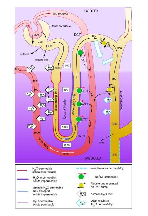
c. Tubular Reabsorption & Secretion
1. Proximal Convoluted Tubule
a. Permeable to solutes (glucose,amino acids,organic molecules, Na+, Ca2+, Cl–, Vitamins);
They are reabsorbed (active)
b. Permeable to water; it’s reabsorbed
(passive), 65% decrease in volume
c. Tubular secretion [H+, K+]
2. Descending limb of loop of nephron
a. Relatively impermeable to solutes
b. Permeable to water, it’s reabsorbed
c. Interstitial gradient promotes reabsorption
3. Loop of nephron (permeable to urea)Urea is recycled
4. Ascending limb of loop of nephron
a. Permeable to NaCl; it’s reabsorbed
b. Impermeable to water
c. Note countercurrent arrangement with 2 parallel tubes running in opposite direction
5. Distal convoluted tubule
a. Aldosterone & ADH
b. Secretion (H+, NH3, drugs)
c. Juxtaglomerular apparatus (renin & erythropoietin precursor)
6. Collecting tubule/ducts
a. Aldosterone causes sodium reabsorption
b. ADH causes water reabsorption
c. Urea
d. Note countercurrent arrangement
d. Vasa recta
Countercurrent thin walled blood vessels serving tubules
1. Maintains interstitial concentrations
2. Aids in concentrating urine
3. Absorbs some water
Nephron Physiology:
10. Other Renal functions
a. Vitamin D activations
b. Erythropoietin precursor
c. Blood Pressure via renin
11. Minimum urine volumes (.5 l/day – 600 mosmol waste/day with maximal concentration of 1200 mosmol/liter)
C. Ureter
Kidney to urinary bladder
D. Urinary bladder
Holds urine (up to 800 mls) in pelvic cavity
– Micturition
Voiding urine – reflex involves stretch of bladder wall causing reflex to spinal cord and back along parasympathetic neurons to contract bladder wall and sphincter relaxes, but control involves impulses from brain which can voluntarily control sphincter
E. Urethra
– Orifice