
HUMAN ANATOMY & PHYSIOLOGY II
Laboratory Objectives Fall 2019
Instructor: Dr. Clare Hays, SI 2032; 303-615-0777, e-mail – [email protected], URL http://sites.msudenver.edu/haysc
Books and Supplies:
- Required: Your textbook is for online or lecture but doesn’t need to come to school with you: Seeley’s Anatomy & Physiology, 12th Ed.,by Van Putte, Regan and Russo including access to Mc-Graw Hill Connect;

2.Required: Your lab manual needs to come to lab with you: Human Anatomy and Physiology Laboratory Manual, 12th Ed., Elaine N. Marieb 
- Optional: Dissection Guide and Atlas to the Mink, by David Smith and Michael Schenk, Morton Publishing;
4. Required: BIO 2320 Dissecting Tools. Available in bookstore; includes a scalpel with replaceable blades, a blunt probe, and small scissors;
5. Not required, but strongly recommended, is a lab coat or an old shirt to protect your clothing. Respirators with filters and eye goggles are available upon request.
Upon completion of lab exercises, you should review the material and do the review sheets from your lab manual, as there are no open lab hours. Lab exams are NOT comprehensive.
MASTERINGAANDP.COM: Your lab manual has some excellent resources for both lecture and lab. These resources and the access code are described at the beginning of your lab manual. You will need to complete a registration process to use this site by clicking you are a student. Then, click Register for Self-Study Access Only and “Mastering is not required for my course.” Enter your access code and click on your book. Go to the Study Area, especially note the PAL section on anatomy.
AUGUST 23 – LANGUAGE OF ANATOMY AND ENDOCRINE ANATOMY
LANGUAGE OF ANATOMY:
Exercise 1: Glance at Figure 1.2 to understand anatomic terminology of the quadriped (mink). Use your own body and the human torso models to refresh on the basic organization of the body. Know the following terms:
Anatomical Position (both human and quadriped), Superior, Inferior, Cranial, Caudal, Medial, Lateral, Superficial, Deep, Ventral, Anterior, Dorsal, Posterior, Proximal, Distal, Sagittal plane, Transverse plane, Frontal plane, Thoracic cavity, Abdominal cavity, Pelvic cavity.
ENDOCRINE ANATOMY:
Exercise 27: Figure 27.1 in the Marieb lab manual has human endocrine pictures and microscopic anatomy. The optional Dissection Guide and Atlas to the Mink by David Smith and Michael Schenk has mink endocrine glands in Chapter 9. Pages 49-50 of optional Mink manual have instructions for opening the ventral body cavity.
- https://www.youtube.com/watch?v=i4IOcct1sOA (Mink opening the throat region)
- https://www.youtube.com/watch?v=KhcPczm08oc (Mink opening thoracic cavity)
- https://www.youtube.com/watch?v=7NEblSo4RrY (Mink opening the abdominal cavity)
- https://www.youtube.com/watch?v=vewQHt-XXX8 (Mink Organ Review)
- https://www.youtube.com/watch?v=07sqb6bdyv4 (Mink Endocrine and Circulatory Preview)
Mink Dissection: Obtain a mink and open the ventral body cavity by cutting through linea alba on the abdomen and then using scissors to cut through the ribcage on the ventral side of the mink, but slightly off center. Be careful not to remove the gonads and do not cut through blood vessels containing colored latex, without first checking with your instructor. Additionally, a fatty greater omentum covers all of the abdominal visceral. You may peel it out of your way, but do not remove it.
- Observe the following endocrine glands of the mink: Thyroid, Thymus, Pancreas, Adrenal, Ovary, Testis. Mink Photographs: Endocrine system mink
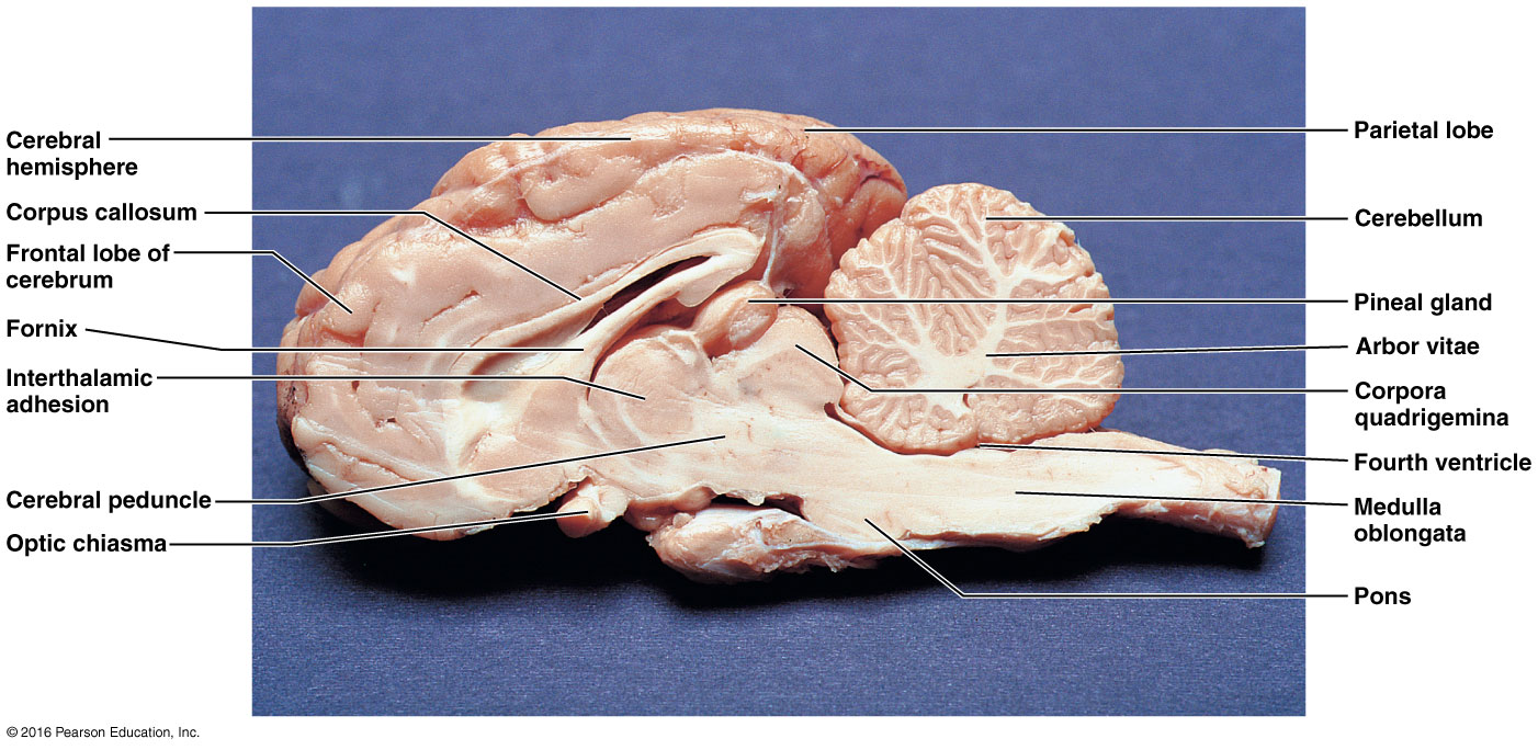
- Observe the pituitary gland and pineal gland (=body) on the preserved sheep brain. See Figures 27.1 and 27.2. Sheep pineal picture in transverse fissure. Sheep pineal picture from sagittal view.
- Put your mink away as described by your instructor. Clean your working area thoroughly.
- Observe the microscopic anatomy on the Thyroid gland, Pancreas, Adrenal gland, Ovary, and Testis as described in Exercise 27 – Activity 2 of lab manual as well as Figures 42.2 and 43.6.
AUGUST 30 – BLOOD
Exercise 29: Complete the following activities using sheep blood or fake blood.
Activity 1: Observe the color and clarity of plasma after you conduct the hematocrit test (to be done later in this lab).
Activity 2: Observe one of each formed elements on a prepared human blood sample slide. You must be able to identify erythrocytes, thrombocytes, and each of the granulocytes (neutrophils, eosinophils, basophils) and each of the agranulocytes (lymphocytes and monocytes). Note: All granulocytes and agranulocytes are types of leukocytes.
Activity 4: Conduct a Hematocrit using the microhematocrit reader card. Then, observe the color and clarity of plasma from Activity 1. See if you can spot the layer of leukocytes found in the buffy coat between the plasma and the red blood cells.
Activity 5: Determine the approximate hemoglobin concentration of the blood sample using the Tallquist method.
Activity 7: Obtain an unknown blood sample and conduct the blood typing experiment to determine its ABO and Rh factor.
SEPTEMBER 6 – ANATOMY OF THE HEART
Exercise 30: Use Figures 30.2, 30.3, 30.4, 30.7, 30.8 for your heart anatomy.
Note: the following URL has good sheep heart pictures: https://homes.bio.psu.edu/faculty/strauss/anatomy/
Observe the sheep heart which has been cut in a frontal section. You are responsible for the following structures:
Visceral pericardium (epicardium), myocardium, endocardium, coronary blood vessels, left and right atria, left and right ventricles, auricles, pulmonary trunk, aorta, aortic semilunar valve, pulmonary veins, superior and inferior vena cavae, right atrioventricular valve (tricuspid), pulmonary semilunar valve, interventricular septum, papillary muscles, chordae tendineae, and left atrioventricular valve (bicuspid).
Mediastinum, pericardial sac, and pericardial cavity are best observed on your mink. See pictures 3 & 4, mediastinum and pericardial sac: CV upper vessels mink
Observe the microscopic anatomy of cardiac muscle as described in Exercise 30 – Activity 4 and Figure 30.6.
SEPTEMBER 13 – EXAM 1 (25 questions, 50 points)
SEPTEMBER 20, 27 – BLOOD VESSELS
Mink blood vessels may be found in Chapter 5 of the optional Mink lab manual or at the following links. Mink blood vessel photographs: CV upper vessels mink Hepatic portal system mink CV- lower vessels mink
- Cat videos who have similar vessels to the mink
- https://www.youtube.com/watch?v=2BAUSMJo7TQ (Cat Circulatory System. Very nicely dissected vessels. Anterior Mesenteric= Superior Mesenteric, Posterior Mesenteric=Inferior Mesenteric)
- https://www.youtube.com/watch?v=3jHnHGn5axw (Mink Lower Vessels)
Dissect your mink and locate the following blood vessels:
Vessels Cranial to Diaphragm: Coronary vessels, superior vena cava (=cranial vena cava = precava), inferior vena cava (=caudal vena cava = postcava), pulmonary trunk (arteries), pulmonary veins, aorta.
Brachiocephalic veins, external jugular veins, internal jugular veins, subclavian veins, axillary veins, brachial veins.
Right brachiocephalic artery, left subclavian artery, right subclavian artery, common carotid arteries, axillary arteries, brachial arteries. (note: both common carotid arteries come off of the brachiocephalic artery in minks, but in humans, the left common carotid artery actually comes off of the aortic arch)
Vessels Caudal to Diaphragm: Adrenolumbar=Suprarenal veins, renal veins, testicular or ovarian veins, iliolumbar veins, common iliac veins, internal iliac veins, external iliac veins, femoral veins.
Hepatic portal vein, gastrosplenic vein, superior mesenteric vein (=cranial mesenteric vein), inferior mesenteric vein (=caudal mesenteric vein). (These vessels are prepared with yellow dye.)
Aorta, celiac trunk, left gastric artery, hepatic artery, splenic artery, superior mesenteric artery (=cranial mesenteric artery), adrenolumbar= suprarenal arteries, renal arteries, testicular or ovarian arteries, inferior mesenteric artery (=caudal mesenteric artery), iliolumbar arteries, external iliac arteries (note: minks do not have a common iliac artery as humans do), internal iliac arteries, femoral arteries.
OCTOBER 4 – CARDIOVASCULAR PHYSIOLOGY (Both Exercises 31 and 33)
Exercise 31: Record your electrocardiogram (ECG or EKG) and identify the P wave, QRS complex, and the T wave. Understand what events are taking place during these 3 recognizable waves.
ELECTROCARDIOGRAPHY; USING INTELITOOL CARDIOCOMP TM
GETTING STARTED
1. With PC computer, McADDAM II and ECG cables connected, you may power up the PC and McADDAM II. The power indicator on the upper right hand corner of McADDAM II will light when the unit is turned on.
2. Launch the Cardiocomp program by clicking on the start menu and highlighting programs, Intelitool, and the select Cardiocomp1. Once loaded, pull down file menu, select new, and click ok.
3. The data collection will stop automatically after the time specified under File Setup. You may change the duration to 60 seconds for shorter collection period. If desired, the sample rate may be slowed down to 60 samples/second (measured in Hz) from Rate setting under File Setup.
4. Although all three leads should be examined during this exercise, start with Lead II. Select I, II, or III to change Leads under the Lead pull down menu.
PREPARING THE SUBJECT
1.Snap the three flat plate electrodes to the black, green and red cables.
2.Remove any jewelry on the wrists or ankles. Scrub the area thoroughly where the electrodes are to be applied {See locations on Lead 1,2,3 diagrams on computer.} Wipe the cleaned area with 70% alcohol on cotton.
3.Apply a liberal amount of electrode gel to the contact surface of the electrode.
4.Strap the electrode to the appropriate appendage (anywhere on the wrist and ankle) so the strap is snug but not too tight . Make sure the subject is comfortable and that their circulation is not restricted.
5.In addition to proper electrode application, signal clarity and stability can be enhanced by ensuring that the electrode cables remain stationary during data acquisition. Any wiggling of the wires relative to each other will introduce noise into the signal. Try to keep the wires being used in a group – taping them together often helps. Also, do not drape any of the wires, including the one which plugs into the computer, over a potential noise source such as a power cord. If the subject is in a supine position on a table, it will be easier to keep the wires stationary.
6.Remember to move cables to the appropriate appendage on the subject when changing Leads on the computer! The ground electrode must be used on the right leg at all times.
THE ELECTROCARDIOGRAM
1.When the subject is ready, click “Start” to begin acquiring data. You can stop data acquisition at any time by clicking the mouse.
2.Examine and analyze your data. Identify the P wave, QRS complex, and T wave. Note any differences in the appearance of the various waves for the different Leads.
3.Select Analyze from under the Acquire pull down menu or by clicking the shortcut button located on the top-right of the Data Acquisition window. Time/Voltage allows you to measure time intervals and voltage differences between any two points in the data set. The numbers displayed in the “Difference” box represent the difference between two data points identified by data markers. Create data markers by placing arrow on desired data point on ECG and clicking mouse. If you hold the mouse button and drag the mouse after creating the first data marker, you can see the absolute voltage for any point in the window. Click “Reset” to erase data markers so that they can be positioned elsewhere in the data set. An automatic analysis may be used by selecting the on field to replace using data markers.
INTERPRETATION OF ELECTROCARDIOGRAM
–The P wave represents atrial depolarization and the QRS complex represents ventricular depolarization. The T wave represents ventricular repolarization. Atrial repolarization is not visible, as it occurs during the dominant QRS complex.
–The P-Q interval is often called the P-R interval because the Q wave is usually small or absent. The normal P-Q interval time is 0.12-0.20 sec. The normal QRS complex duration is less than 0.10 sec, and the Q-T interval should be less than 0.38 sec. How do your results compare with these normal values?
–Determine the ventricular rate by measuring the elapsed time between two R waves. Divide 60 by your time between R waves. The ventricular rate is in beats per minute.
Exercise 33: Complete the following activities for heart sounds, blood pressure and pulse determinations.
Activity 1: Complete Auscultating Heart Sounds.
Activity 2: Palpate Superficial Pulse Points.
Activity 4: Taking an Apical Pulse.
Activity 5: Use a sphygmomanometer to measure arterial blood pressure.
Activity 6: Estimate your venous pressure, both at rest and while performing the Valsalva maneuver.
Activity 7: Observe the effect of posture, exercise and ice water on blood pressure and heart rate.
Activity 8: Observe the effect of local chemical and physical factors on skin color.
OCTOBER 11 – EXAM 2
OCTOBER 18 – ANATOMY OF RESPIRATORY AND DIGESTIVE SYSTEMS
RESPIRATORY ANATOMY:
Exercise 36: Complete histology and mink respiratory dissection. Chapter 6 of your optional Mink manual has respiratory anatomy.
- Examine a microscopic section of lung tissue. See Figure 36.7.
- Dissect your mink and find the following:
External nares, trachea, larynx, epiglottis, glottis, hyoid bone, vagus nerve, primary bronchi, pleural cavity, parietal pleura, visceral pleura, diaphragm, and lungs.
Mink photos of the respiratory system
https://www.youtube.com/watch?v=pdhQ7kIwXiQ (Mink Respiratory System)
DIGESTIVE ANATOMY:
Exercise 38: Complete histology and mink digestive dissection. Chapter 4 of your optional Mink manual has digestive anatomy.
- Observe microscopic sections of the stomach, small intestine, pancreas, liver, colon and taste buds. See Figures 38.6, 38.9, 38.16, 26.3, 27.3c.
- Dissect your mink and find the following:
Parotid salivary gland and duct, teeth, oral cavity, hard palate, tongue papillae, frenulum of tongue, esophagus, peritoneal cavity, parietal peritoneum, visceral peritoneum,liver, greater omentum, gall bladder, stomach [cardia, fundus, body, pylorus], greater and lesser curvature of stomach, lesser omentum, pancreas, spleen, common bile duct, small intestine [duodenum, jejunum, ileum], mesentery proper, large intestine including: colon [ascending, transverse, descending], rectum, anus.
Mink photos of the digestive system
https://www.youtube.com/watch?v=cNpspeKmCjQ (Mink Digestive System with explanation)
https://www.youtube.com/watch?v=YQWIxAQbM_c (Mink Digestive System Review)
OCTOBER 25 – Exercises 43& 44: No on-campus meeting. Complete the review sheet assignment which consists of completing 1. the Review Sheet in your Marieb Laboratory Manual “Exercise 43 Review Sheet: Physiology of Human Reproduction: Gametogenesis and the Female Cycles” PLUS 2. Review Sheet in your Marieb Laboratory Manual “Exercise 44 Review Sheet: Survey of Embryonic Development.” You may do these review sheets at home. The two review exercises are due when November 1 when you come in for your next lab. You may hand these in to me during lab, or scan/photograph them and submit them electronically. You will lose 5 points per day that they are submitted late. 10 points are possible for complete and accurate answers of each review exercise for a total of 20 points.
NOVEMBER 1 – RESPIRATORY PHYSIOLOGY
Exercise 37: Complete the following physiology activities.
Activity 2: Auscultating Respiratory Sounds using a stethoscope.
Activity 3: Use a wet spirometer or the Vernier Probes with attached spirometer to record your respiratory volumes and capacities.
Record the following: Respirations per minute, Tidal Volume, Minute Respiratory Volume, Expiratory Reserve Volume, Vital Capacity, and Inspiration Reserve Volume
Instructions for using the Vernier Sensors: -Connect the Vernier Spirometer to the LabQuest interface, channel 1.
-Assemble the mouthpieces by placing the larger diameter side of a white bacterial filter to the “Inlet” side of the Spirometer. Attach a gray disposable mouthpiece to the other end of the bacterial filter. The assembly is similar to Figure 37A.12.
-Hold the Spirometer assembly in both hands and brace your arms against the table. Make sure that you hold the spirometer straight up and down and do not move it during data collection. Click on Sensor and choose “zero” for calibration.
-Click the “play” button to begin data collection. Record the Tidal Volume, Inspiratory Reserve Volume, Expiratory Reserve Volume and Vital Capacity as described in your lab manual in Activity 3. Inspiratory values go downward on the screen and expiratory values go up. Click on the peak of your waves and record the flow rate.
3. Activity 6: Complete this activity without using a spirometer.
NOVEMBER 8 – ANATOMY OF URINARY AND REPRODUCTIVE SYSTEMS
Your optional Mink lab manual has urinary and reproduction in Chapter 7. Mink photographs of urinary and reproductive anatomy: Reproductive system mink
- https://www.youtube.com/watch?v=Hz9Q2GMy-HQ (Mink Digestion Review, Vessel Review, Urinary System Review & Male Reproductive System)
- https://www.youtube.com/watch?v=PtBCx9RmNOI (Mink Female Reproductive System)
Dissect your mink. You are responsible for the following:
Kidney, renal capsule, renal cortex, renal medulla, renal pelvis, hilus, ureter, urinary bladder, and urethra.
Penis, testes, spermatic cord, ductus deferens (=vas deferens), inguinal canal, prostate gland.
Uterus (uterine body & 2 uterine horns), uterine tube (=oviduct=fallopian tube), ovary, vagina, cervix, and vulva.
Observe the microscopic anatomy of the ovary and testis. Refer to Figures 42.2 and 43.6.
NOVEMBER 15 – URINALYSIS & REPRODUCTIVE PHYSIOLOGY – (Attendance will be taken)
Exercise 41: Analyze a urine sample from your own urine and from an unknown provided. Interpret your results.
Activity 1: Complete as much of the Urinalysis Results form as possible using information derived from observation, reagent strips, and the refractometer.
Activity 2: Complete a sediment analysis on your own urine as described.
A one-hour film on reproduction will be viewed during your laboratory period.