
Books and Supplies:
- Required: Your textbook is for online or lecture, but doesn’t need to come to school with you: Seeley’s Anatomy & Physiology, 13th Ed., by Van Putte, Regan and Russo including access to Mc-Graw Hill Connect;
2.Required: Your lab manual needs to come to lab with you: Human Anatomy and Physiology Laboratory Manual, 12th Ed., Elaine N. Marieb 
- Optional: Dissection Guide and Atlas to the Mink, by David Smith and Michael Schenk, Morton Publishing;
4. Required: BIO 2310 Dissecting Tools. Available in bookstore; includes a scalpel with replaceable blades, a blunt probe, and small scissors;
5. Not required, but strongly recommended, is a lab coat or an old shirt to protect your clothing. Respirators with filters and eye goggles are available upon request.
6. Familiarize yourself with the safety rules for lab and dissection protocols.
Upon completion of lab exercises, you should review the material and do the review sheets from your lab manual, as there are no open lab hours. Lab exams are NOT comprehensive.
MASTERINGAANDP.COM: Your lab manual has some excellent resources for both lecture and lab. These resources and the access code are described at the beginning of your lab manual. You will need to complete a registration process to use this site by clicking you are a student. Then, click Register for Self-Study Access Only and “Mastering is not required for my course.” Enter your access code and click on your book. Go to the Study Area, especially note the PAL section on anatomy.
Instructor Information: Dr. Hays [email protected]
Office hours Tuesdays 8-1 in Science 2032 or on Teams
EXTRA CREDIT OPPORTUNITY: Attending lab is essential to success in the class, as it provides visual and tactile input to the structures you need to know as well as relationships of one structure to another. You may earn extra credit points for attending labs throughout the semester as follows, provided that you are not more than 20 minutes late to any given lab:
Attending fewer than 3 labs: 0 points extra credit
Attending 3-4 labs: 5 points extra credit
Attending 5 labs: 10 points extra credit
Attending 6-7 labs: 15 points extra credit
Attending 8-9 labs: 20 points extra credit
Attending all 10 in-person labs: 25 points extra credit
JANUARY 19 – LANGUAGE OF ANATOMY AND ENDOCRINE ANATOMY
LANGUAGE OF ANATOMY:
Exercise 1: Glance at Figure 1.2 to understand anatomic terminology of the quadruped (mink). Use your own body and the human torso models to refresh on the basic organization of the body. Know the following terms:
Anatomical Position (both human and quadruped), Superior, Inferior, Cranial, Caudal, Medial, Lateral, Superficial, Deep, Ventral, Anterior, Dorsal, Posterior, Proximal, Distal, Sagittal plane, Transverse plane, Frontal plane, Thoracic cavity, Abdominal cavity, Pelvic cavity.
ENDOCRINE ANATOMY:
Exercise 27: Figure 27.1 in the Marieb lab manual has human endocrine pictures and microscopic anatomy. The optional Dissection Guide and Atlas to the Mink by David Smith and Michael Schenk has mink endocrine glands in Chapter 9. Pages 49-50 of optional Mink manual have instructions for opening the ventral body cavity.
- https://www.youtube.com/watch?v=i4IOcct1sOA (Mink opening the throat region)
- https://www.youtube.com/watch?v=KhcPczm08oc (Mink opening thoracic cavity)
- https://www.youtube.com/watch?v=7NEblSo4RrY (Mink opening the abdominal cavity)
- https://www.youtube.com/watch?v=vewQHt-XXX8 (Mink Organ Review)
- https://www.youtube.com/watch?v=07sqb6bdyv4 (Mink Endocrine and Circulatory Preview)
Mink Dissection: Obtain a mink and open the ventral body cavity by cutting through linea alba on the abdomen and then using scissors to cut through the ribcage on the ventral side of the mink, but slightly off center. Be careful not to remove the gonads and do not cut through blood vessels containing colored latex, without first checking with your instructor. Additionally, a fatty greater omentum covers all of the abdominal visceral. You may peel it out of your way, but do not remove it.
- Observe the following endocrine glands of the mink: Thyroid, Thymus, Pancreas, Adrenal, Ovary, Testis. Mink Photographs: Endocrine system mink
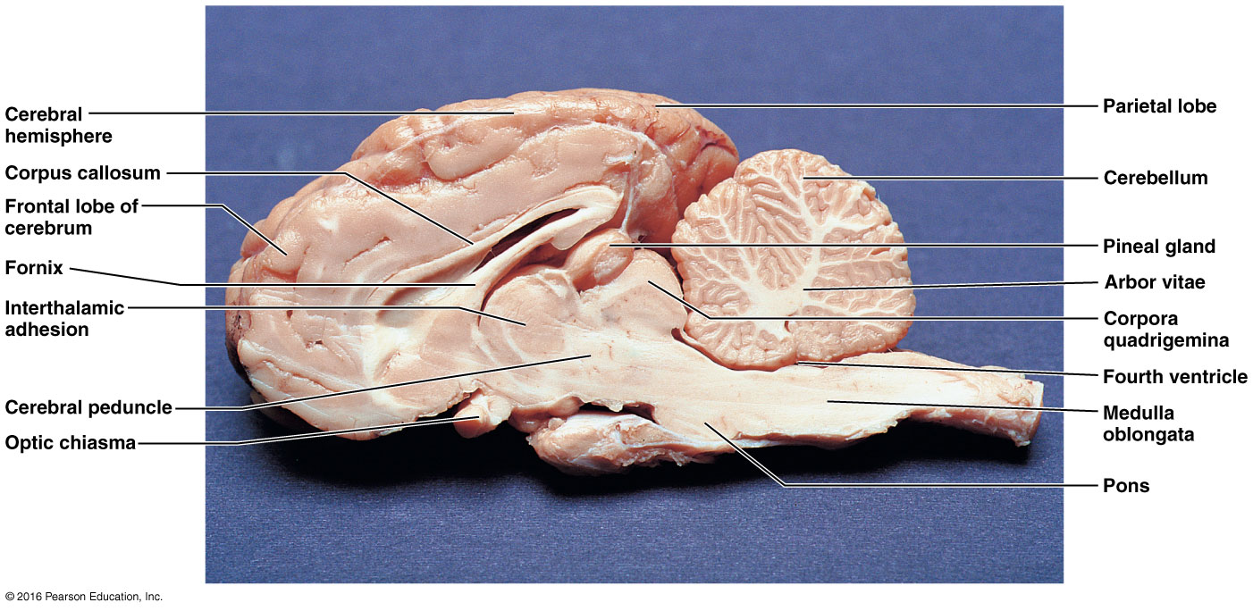
- Observe the pituitary gland and pineal gland (=body) on the preserved sheep brain. See Figures 27.1 and 27.2. Sheep pineal picture in transverse fissure. Sheep pineal picture from sagittal view.
- Put your mink away as described by your instructor. Clean your working area thoroughly.
- Observe the microscopic anatomy on the Thyroid gland, Pancreas, Adrenal gland, Ovary, and Testis as described in Exercise 27 – Activity 2 of lab manual as well as Figures 42.2 and 43.6.
JANUARY 26 – BLOOD
Exercise 29: Complete the following activities using sheep blood or fake blood.
Activity 1: Observe the color and clarity of plasma after you conduct the hematocrit test (to be done later in this lab).
Activity 2: Observe one of each formed elements on a prepared human blood sample slide. You must be able to identify erythrocytes, thrombocytes, and each of the granulocytes (neutrophils, eosinophils, basophils) and each of the agranulocytes (lymphocytes and monocytes). Note: All granulocytes and agranulocytes are types of leukocytes.
Activity 4: Conduct a Hematocrit using the microhematocrit reader card. Then, observe the color and clarity of plasma from Activity 1. See if you can spot the layer of leukocytes found in the buffy coat between the plasma and the red blood cells.
Activity 5: Determine the approximate hemoglobin concentration of the blood sample using the Tallquist method.
Activity 7: Obtain an unknown blood sample and conduct the blood typing experiment to determine its ABO and Rh factor.
FEBRUARY 2 – ANATOMY OF THE HEART
Exercise 30: Use Figures 30.2, 30.3, 30.4, 30.7, 30.8 for your heart anatomy.
Observe the sheep heart which has been cut in a frontal section. You are responsible for the following structures:
Visceral pericardium (epicardium), myocardium, endocardium, coronary blood vessels, left and right atria, left and right ventricles, auricles, pulmonary trunk, aorta, aortic semilunar valve, pulmonary veins, superior and inferior vena cavae, right atrioventricular valve (tricuspid), pulmonary semilunar valve, interventricular septum, papillary muscles, chordae tendineae, and left atrioventricular valve (bicuspid).
Mediastinum, pericardial sac, and pericardial cavity are best observed on your mink. See pictures 3 & 4, mediastinum and pericardial sac: CV upper vessels mink
Observe the microscopic anatomy of cardiac muscle as described in Exercise 30 – Activity 4 and Figure 30.6.
Here is a good heart overview video made by Sophie, a previous TA.
FEBRUARY 8-10 LAB EXAM ONE You do not come to lab this week, as the exam is on Canvas. The online lab test is primarily fill in the blank questions randomly selected from a test bank and spelling must be exact. It may be found on MSU Denver Canvas. It opens Thursday Feb 8 at 12 am and closes at 11:59 pm on Saturday Feb 10. The online tests have 25 questions at 2 points each, 30 minutes. The exam will only test you on material from these laboratory objectives. I do put my eyes on everyone’s completed exams to double check the computer grading.
Read Exam Directions:
- All answers for fill-in-the-blank questions are in lower case letters and must be spelled correctly.
- You never have to indicate right or left unless it is a heart chamber.
- You will need to specify artery or vein unless that word is given in the question.
Any lab exam not taken by 11:59 pm on Saturday Feb 10 will have a 10% deduction per day late.
FEBRUARY 16 and 23 – BLOOD VESSELS
Mink blood vessels may be found in Chapter 5 of the optional Mink lab manual or at the following links. Mink blood vessel photographs: CV upper vessels mink Hepatic portal system mink CV- lower vessels mink
- Cat videos who have similar vessels to the mink
- https://www.youtube.com/watch?v=2BAUSMJo7TQ (Cat Circulatory System. Very nicely dissected vessels. Anterior Mesenteric= Superior Mesenteric, Posterior Mesenteric=Inferior Mesenteric)
- https://www.youtube.com/watch?v=3jHnHGn5axw (Mink Lower Vessels)
Dissect your mink and locate the following blood vessels:
Vessels Cranial to Diaphragm: Coronary vessels, superior vena cava (=cranial vena cava = precava), inferior vena cava (=caudal vena cava = postcava), pulmonary trunk (arteries), pulmonary veins, aorta.
Brachiocephalic veins, external jugular veins, internal jugular veins, subclavian veins, axillary veins, brachial veins.
Right brachiocephalic artery, left subclavian artery, right subclavian artery, common carotid arteries, axillary arteries, brachial arteries. (note: both common carotid arteries come off of the brachiocephalic artery in minks, but in humans, the left common carotid artery actually comes off of the aortic arch)
Vessels Caudal to Diaphragm: Adrenolumbar=Suprarenal veins, renal veins, testicular or ovarian veins, iliolumbar veins, common iliac veins, internal iliac veins, external iliac veins, femoral veins.
Hepatic portal vein, gastrosplenic vein, superior mesenteric vein (=cranial mesenteric vein), inferior mesenteric vein (=caudal mesenteric vein). (These vessels are prepared with yellow dye.)
Aorta, celiac trunk, left gastric artery, hepatic artery, splenic artery, superior mesenteric artery (=cranial mesenteric artery), adrenolumbar= suprarenal arteries, renal arteries, testicular or ovarian arteries, inferior mesenteric artery (=caudal mesenteric artery), iliolumbar arteries, external iliac arteries (note: minks do not have a common iliac artery as humans do), internal iliac arteries, femoral arteries.
MARCH 1 – CARDIOVASCULAR PHYSIOLOGY (Both Exercises 31 and 33)
Exercise 31: Understand what is recorded with an electrocardiogram (ECG or EKG) and where leads are placed for a Lead II EKG. On the Lead II EKG, identify the P wave, QRS complex, and the T wave. Understand what events are taking place during these 3 recognizable waves. Observe abnormal EKGs and the important information that can be gleaned on heart pathology from these. However, you will only be tested on normal EKGs.
INTERPRETATION OF ELECTROCARDIOGRAM: The P wave represents atrial depolarization and the QRS complex represents ventricular depolarization. The T wave represents ventricular repolarization. Atrial repolarization is not visible, as it occurs during the dominant QRS complex.
Exercise 33: Complete the following activities for heart sounds, blood pressure and pulse determinations.
Activity 1: Complete Auscultating Heart Sounds.
Activity 2: Palpate Superficial Pulse Points.
Activity 4: Taking an Apical Pulse.
Activity 5: Use a sphygmomanometer to measure arterial blood pressure.
Activity 6: Estimate your venous pressure, both at rest and while performing the Valsalva maneuver.
Activity 7: Observe the effect of posture, exercise and ice water on blood pressure and heart rate.
Activity 8: Observe the effect of local chemical and physical factors on skin color.
MARCH 7-9 LAB EXAM TWO You do not come to lab this week, as the exam is on Canvas. The online lab test is primarily fill in the blank questions randomly selected from a test bank and spelling must be exact. It may be found on MSU Denver Canvas. It opens Thursday Mar 7 at 12 am and closes at 11:59 pm on Saturday March 9. The online tests have 25 questions at 2 points each, 30 minutes. The exam will only test you on material from these laboratory objectives. I do put my eyes on everyone’s completed exams to double check the computer grading.
Read Exam Directions:
- All answers for fill-in-the-blank questions are in lower case letters and must be spelled correctly.
- You never have to indicate right or left, but you do need to specify artery or vein unless it is given in the question.
Any lab exam not taken by 11:59 pm on Saturday Mar 9 will have a 10% deduction per day late.
MARCH 15 – ANATOMY OF RESPIRATORY AND DIGESTIVE SYSTEMS
RESPIRATORY ANATOMY:
Exercise 36: Complete histology and mink respiratory dissection. Chapter 6 of your optional Mink manual has respiratory anatomy.
- Examine a microscopic section of lung tissue. See Figure 36.7.
- Dissect your mink and find the following:
External nares, trachea, larynx, epiglottis, glottis, hyoid bone, vagus nerve, primary bronchi, pleural cavity, parietal pleura, visceral pleura, diaphragm, and lungs.
Mink photos of the respiratory system
https://www.youtube.com/watch?v=pdhQ7kIwXiQ (Mink Respiratory System)
DIGESTIVE ANATOMY:
Exercise 38: Complete histology and mink digestive dissection. Chapter 4 of your optional Mink manual has digestive anatomy.
- Observe microscopic sections of the stomach, small intestine, pancreas, liver, colon and taste buds. See Figures 38.6, 38.9, 38.16, 26.3, 27.3c.
- Dissect your mink and find the following:
Parotid salivary gland and duct, teeth, oral cavity, hard palate, tongue papillae, frenulum of tongue, esophagus, peritoneal cavity, parietal peritoneum, visceral peritoneum, liver, greater omentum, gall bladder, stomach [cardia, fundus, body, pylorus], greater and lesser curvature of stomach, lesser omentum, pancreas, spleen, common bile duct, small intestine [duodenum, jejunum, ileum], mesentery proper, large intestine including: colon [ascending, transverse, descending], rectum, anus.
Mink photos of the digestive system
https://www.youtube.com/watch?v=cNpspeKmCjQ (Mink Digestive System with explanation)
https://www.youtube.com/watch?v=YQWIxAQbM_c (Mink Digestive System Review)
MARCH 29 – Exercises 37, 45: No on-campus meeting. Use this time at home to complete 4 required virtual labs on McGraw Hill Connect. There will be test questions from these labs on Lab Exam 3. You may do the labs as many times as you wish, and only your BEST score will be delivered to your instructor.
1. Virtual Lab Tutorial. 10 points possible. Due by May 11 at 11:59 pm
2. Mechanism of Breathing. 10 points possible. Due by May 11 at 11:59 pm. Here are some study notes from this lab to prepare for lab exam 3: Virtual Lab Note Mechanism of Breathing
3. Pulmonary Function Tests. 10 points possible. Due by May 11 at 11:59 pm. Here are some study notes from this lab to prepare for lab exam 3: Virtual Lab Pulmonary Function
4. Human Genetics: Chromosome Inheritance during Meiosis. 10 points possible. Due by May 11 at 11:59 pm. Here are some study notes from this lab to prepare for lab exam 3: Virtual Lab Notes Genetics and Meiosis
APRIL 5 – RESPIRATORY PHYSIOLOGY
Exercise 37: Complete the following physiology activities.
Activity 2: Auscultating Respiratory Sounds using a stethoscope.
Activity 3: Measuring Respiratory Volumes using a Non-Recording Wet Spirometer
One person from each group should be the subject and complete #1-7 in your lab manual. Measurements should be recorded for the following
- Respiratory Rate (RR)
- Tidal Volume (TV)
- Minute Respiratory Volume (TV X RR)
- Expiratory Reserve Volume (ERV)
- Vital Capacity (VC)
- Inspiratory Reserve Volume (IRV) Use the calculation IRV = VC – (TV + ERV)
Activity 6: Complete this activity without using a spirometer. Determine the main regulator of breathing.
Review the Pulmonary Function Test Virtual Lab Terms
APRIL 12 – ANATOMY OF URINARY AND REPRODUCTIVE SYSTEMS
Your optional Mink lab manual has urinary and reproduction in Chapter 7. Mink photographs of urinary and reproductive anatomy:
Urinary structures of the mink
- https://www.youtube.com/watch?v=Hz9Q2GMy-HQ (Mink Digestion Review, Vessel Review, Urinary System Review & Male Reproductive System)
- https://www.youtube.com/watch?v=PtBCx9RmNOI (Mink Female Reproductive System)
Dissect your mink. You are responsible for the following:
Kidney, renal capsule, renal cortex, renal medulla, renal pelvis, hilus (or hilum), ureter, urinary bladder, and urethra.
Penis, testes, spermatic cord, ductus deferens (=vas deferens), inguinal canal, prostate gland.
Uterus (uterine body & 2 uterine horns), uterine tube (=oviduct=fallopian tube), ovary, vagina, cervix, and vulva.
Observe the microscopic anatomy of the ovary and testis. Refer to Figures 42.2 and 43.6.
APRIL 19 – URINALYSIS & REPRODUCTIVE PHYSIOLOGY
Exercise 41: Analyze a urine sample from your own urine and from an unknown provided. Interpret your results.
Activity 1: Complete as much of the Urinalysis Results form as possible using information derived from observation, reagent strips, and the refractometer.
Activity 2: Complete a sediment analysis on your own urine as described.
A one-hour film on reproduction, Miracle of Life, may be viewed at home. There will be simple exam questions on the film (meaning watch it, but no need to take notes). Here is a link to the film: https://archive.org/details/NOVATheMiracleOfLife
APRIL 25 – 27 LAB EXAM THREE You do not come to lab this week, as the exam is on Canvas. The online lab test is primarily fill in the blank questions randomly selected from a test bank and spelling must be exact. It may be found on MSU Denver Canvas. It opens Thursday Apr 25 at 12 am and closes at 11:59 pm on Saturday April 27. The online tests have 25 questions at 2 points each, 30 minutes. The exam will only test you on material from these laboratory objectives. I do put my eyes on everyone’s completed exams to double check the computer grading.
Read Exam Directions:
- All answers for fill-in-the-blank questions are in lower case letters and must be spelled correctly.
- You never have to indicate right or left.
Any lab exam not taken by 11:59 pm on Saturday Apr 27 will have a 10% deduction per day late.