
Laboratory Objectives for Virtual A&P II Labs Fall 2020
Instructor: Dr. Clare Hays, SI 2032; e-mail – [email protected], URL for website: http://sites.msudenver.edu/haysc
Books and Supplies:
- Required: Your Connect online access comes with this e-book. Seeley’s Anatomy & Physiology, 12th Ed.,by Van Putte, Regan and Russo;

2.Required: Human Anatomy and Physiology Laboratory Manual, 12th Ed., Elaine N. Marieb. 
Note: All Exercise numbers listed below refer to the Marieb Lab Manual.
WEEK 1, AUGUST 17-23 – LANGUAGE OF ANATOMY AND ENDOCRINE ANATOMY
LANGUAGE OF ANATOMY:
Exercise 1 in Marieb: Review understand anatomic terminology. Use your own body and the human torso models to refresh on the basic organization of the body. Know the following terms:
Anatomical Position, Superior, Inferior, Cranial, Caudal, Medial, Lateral, Superficial, Deep, Ventral, Anterior, Dorsal, Posterior, Proximal, Distal, Sagittal plane, Transverse plane, Frontal plane, Thoracic cavity, Abdominal cavity, Pelvic cavity.
Video of Human Body Organization
ENDOCRINE ANATOMY:
Histology: You are responsible for the being able to identify following endocrine glands from a histology photo on an exam. Observe the microscopic anatomy on the Thyroid gland, Pancreas, Adrenal gland, Ovary, and Testis as described in Exercise 27 – Activity 2 of lab manual as well as Figures 42.2 and 43.6.
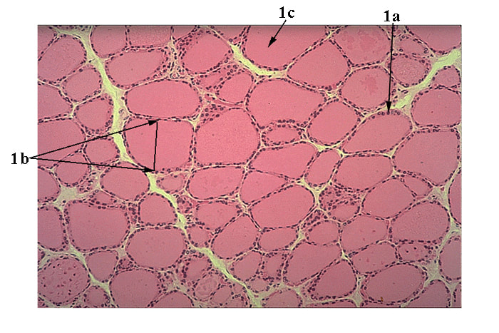
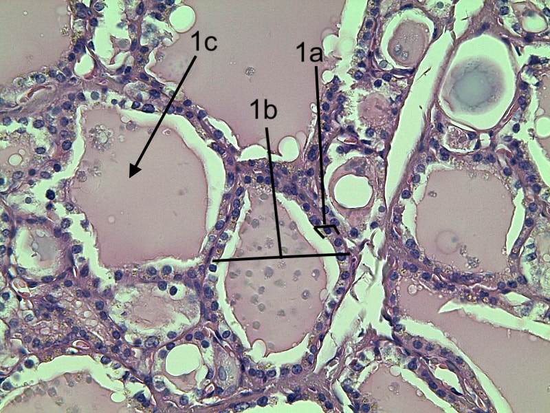
Figure 1: Thyroid gland low power and high power. Note the simple cuboidal epithelium (marked 1a) that surrounds the thyroid follicles. The thyroid follicles are labeled 1b and are filled with colloid (labeled 1c) that is the location of T3 and T4.
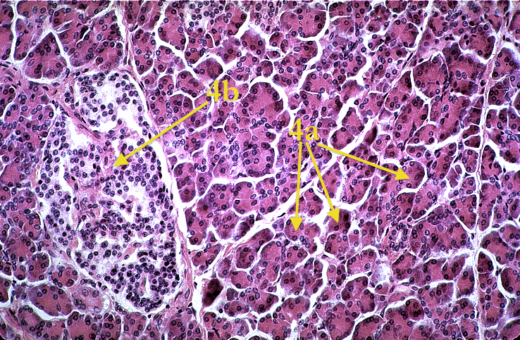
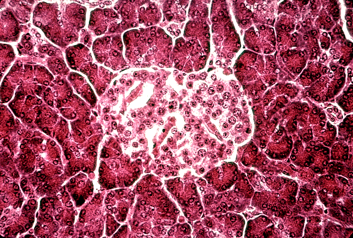
Figure 2: Pancreas gland showing the exocrine acinar cells labeled 4a and the endocrine islets labeled 4b.
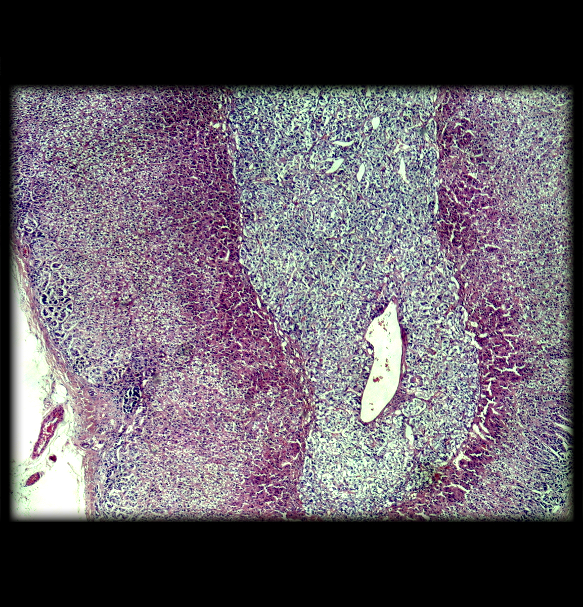
Figure 3: Adrenal gland. Note the layers in the outer adrenal cortex (zona glomerulosa, zona fasciculata, zona reticularis) and the distinctive inner adrenal medulla.
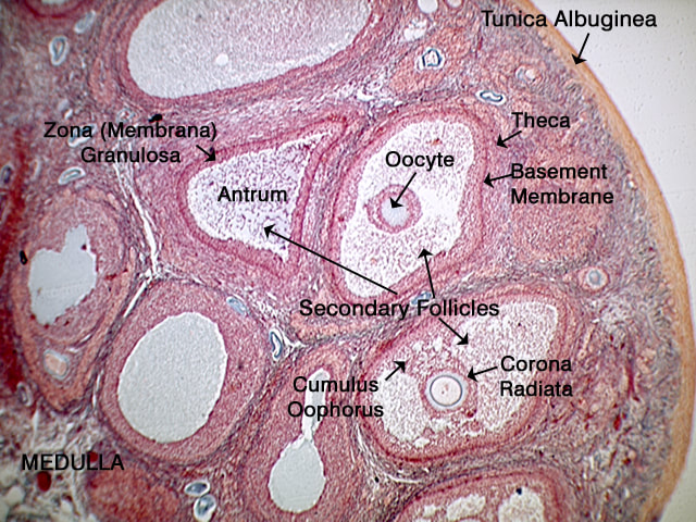
Figure 4: Ovary. Note the various levels of oocyte development within the follicles.
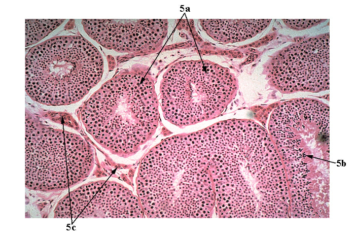
Figure 5: Testes. This is a cross section of the seminiferous tubules in the testes, which is the site of sperm formation. Note that the more immature spermatogonia and spermatocytes are towards the periphery of the tubules. The more mature cells are towards the center lumen of the tubule. In fact if you look closely, you can see the tails of the sperm in the lumen.
Gross Anatomy: Before you find the structures listed below in Anatomy and Physiology Revealed in Connect, watch the help videos so that you are familiar with APR. Once you click on APR, there is a question mark “need help” tab. Click that and then choose “select help video.” I suggest watching the Program Overview, My Course Content, and Dissection help videos. Here is a video on how to use APR: https://myclasses.tegrity.com/#/recording/83b7d082-414d-4624-aea2-100a525fc872?playbackToken=2PB0NHGHYZZNY
You are responsible for the being able to identify following list of endocrine glands from a cadaver photo or model on an exam. Figure 27.1 in the Marieb lab manual has human endocrine pictures. APR in Connect has a specific list of cadaver photos located in Anatomy & Physiology Revealed, My Course Content, List (Endocrine pkrMv) and Module-Endocrine.
Pineal gland, Hypothalamus, Pituitary gland, Thymus, Thyroid gland, Adrenal gland, Pancreas, Parathyroid glands, Gonads (Ovaries and Testes).
Note: There is nothing to hand in or submit when you do the Endocrine lab. In fact, this is the case for many of the labs. The only lab items that will contribute to your grade are the 3 lab exams and the virtual labs that you begin next week.
WEEK 2, AUGUST 24-30 – BLOOD **NOTE LAB ASSIGNMENTS ARE DUE THIS WEEK**
Exercise 29: Blood
You have 4 Virtual Labs to do on Connect. They are all due by August 30 at 11:59 pm. The labs include 1. Virtual Lab Tutorial, 2. Hematocrit Test, 3. Hemoglobin Test, and 4. Blood Typing Test. Each of these labs is worth 10 points for a total of 40 points. Additionally, there will be exam questions from these exercises. Complete the following activities using Virtual Labs (DUE August 30 at 11:59 pm.) and APR. Your lab manual is your reference.
In addition to the 4 virtual labs, you should be able to identify all formed elements found in human blood. You must be able to identify erythrocytes, thrombocytes, and each of the granulocytes (neutrophils, eosinophils, basophils) and each of the agranulocytes (lymphocytes and monocytes). Note: All granulocytes and agranulocytes are types of leukocytes. You can observe all of these in your Marieb lab manual or at APR in Connect. Go to My Course Content, List (Blood e89Jx), Module-Cardiovascular and click on the My Histology link. You may need to turn off the tags and highlighting.
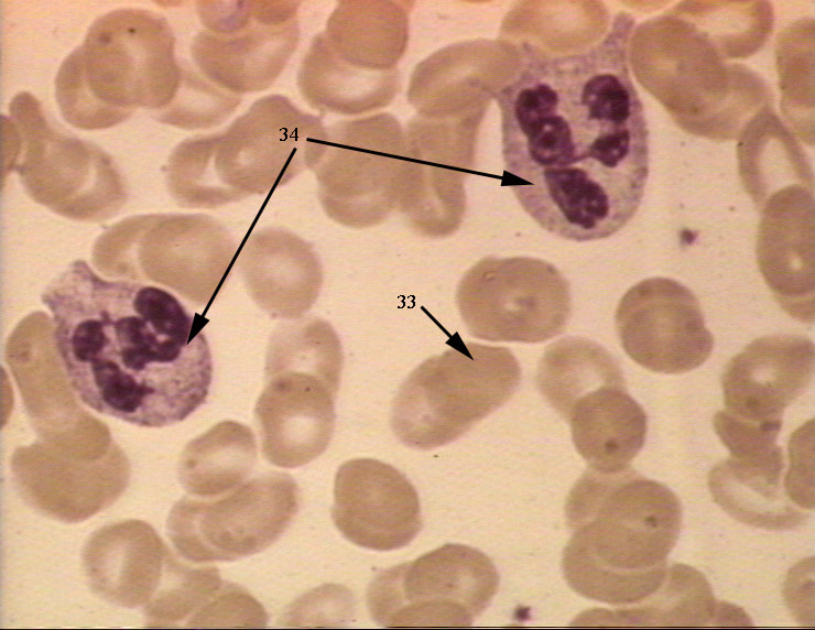


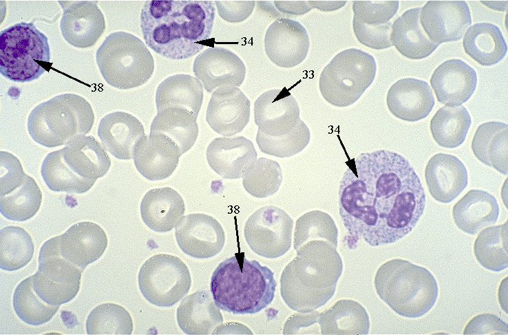
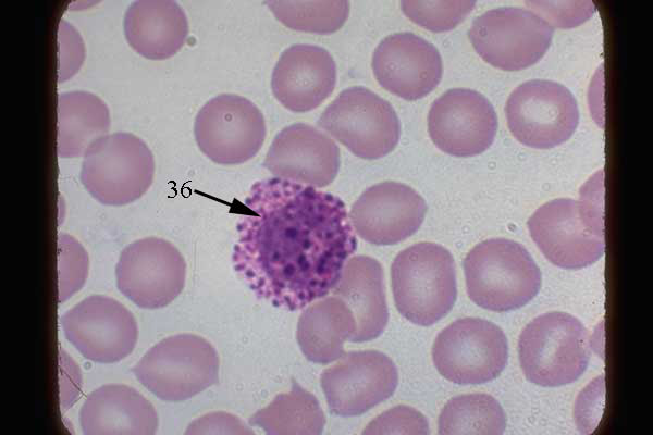
Key to photos: Top left shows erythrocytes (33) and the small dots are thrombocytes, the large cells are neutrophils (34). Top right shows numerous erythrocytes, thrombocytes are the small dots, and the large cell is an eosinophil (35). Middle left shows erythrocytes (33), a couple of thrombocytes, a lymphocyte (38) and a monocyte (37). Middle right shows numerous erythryocytes (33), thrombocytes are the small purple dots, two lymphocytes (38), and two neutrophils (34). Bottom shows erythrocytes and one basophil (36).
Note: Since you cannot go back into your Virtual Labs for studying reasons, the key points from the blood virtual labs may be found here to help you prepare for Lab Exam One: Virtual Lab Blood
WEEK 3, AUGUST 31-SEPTEMBER 6 – ANATOMY OF THE HEART
Exercise 30: Gross Anatomy: You are responsible for the being able to identify following heart structures from a cadaver photo or model on an exam. APR in Connect has a specific list of cadaver photos located in Anatomy & Physiology Revealed, My Course Content, List (Heart PFX9L) and Module-Cardiovascular. Make sure to look at the My Dissection, My Animation, and My Histology. You are responsible for the following gross structures:
Fibrous pericardium, serous pericardium (parietal and visceral layers), myocardium of both ventricles, left and right atria, left and right ventricles, auricles, pulmonary trunk, aorta, aortic semilunar valve, pulmonary veins, superior and inferior vena cavae, right atrioventricular valve (tricuspid), pulmonary semilunar valve, interatrial septum, interventricular septum, papillary muscles, chordae tendineae, and left atrioventricular valve (bicuspid or mitral).
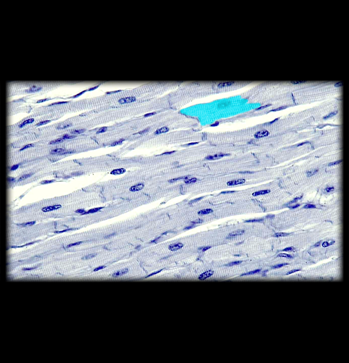
Observe the microscopic anatomy of cardiac muscle as described in Exercise 30 – Activity 4 and Figure 30.6.
WEEK 4: SEPTEMBER 8-13 (at 11:59 pm) – EXAM 1
(25 questions, 50 points)
You will have 45 minutes in one sitting to complete this lab exam online in Connect. You will find it in the Set One Laboratory Exercises & Exams Section. It consists of labeling and multiple choice questions. Read the questions carefully, because a few have multiple answers. There is a 5% deduction in your score for each day you are late taking the exam after the due date.
WEEK 5-6, SEPTEMBER 14-27 – BLOOD VESSELS
Gross Anatomy: You are responsible for the being able to identify following blood vessels from a cadaver photo, diagram, or model on an exam. APR in Connect has a specific list of cadaver photos located in Anatomy & Physiology Revealed, My Course Content, List (Blood Vessels qYh6q) and Module-Cardiovascular. Make sure to look at the My Dissection and My Animation.
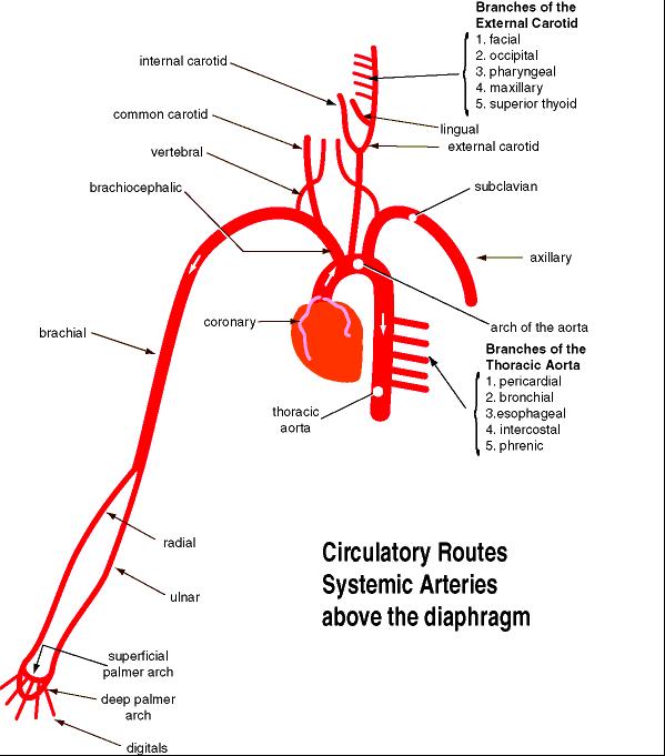
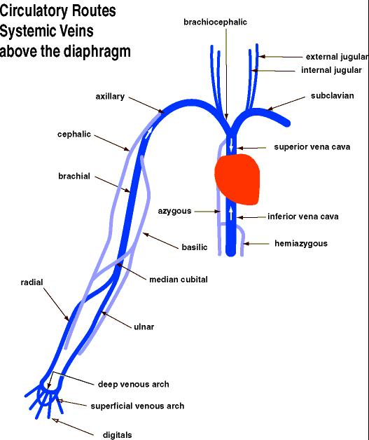
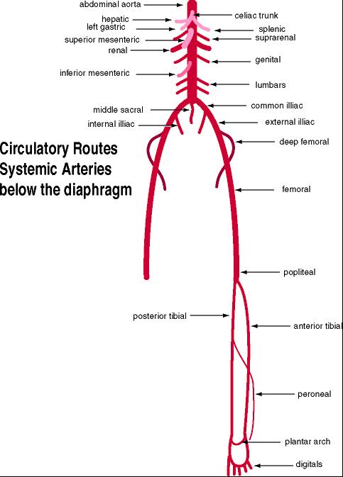
Additionally, these photos are excellent flow charts to understand the blood vessels.
Human arteries above the diaphragm:
Human veins above the diaphragm:
Human arteries below the diaphragm:
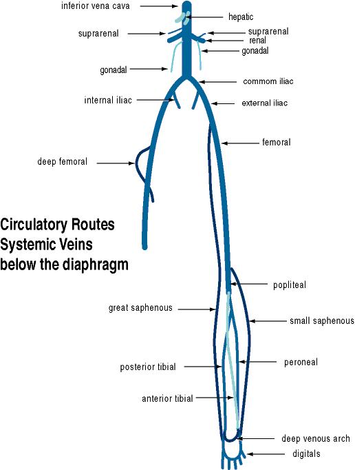
Human veins below the diaphragm:
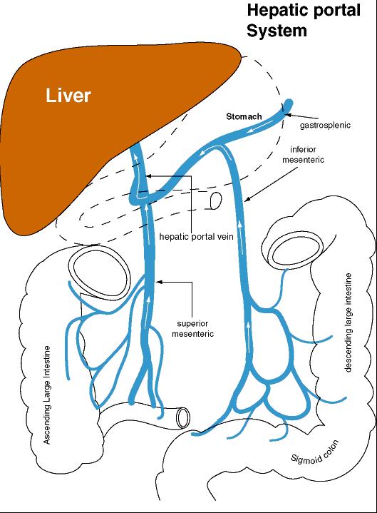
Human Hepatic Portal System:
You are responsible for the following structures:
Vessels Superior to Diaphragm: Superior vena cava, azygos vein, pulmonary trunk (arteries), pulmonary veins, aorta.
Brachiocephalic veins, external jugular veins, internal jugular veins, subclavian veins, axillary veins, brachial veins.
Right brachiocephalic artery (trunk), left subclavian artery, right subclavian artery, common carotid arteries, external carotid arteries, internal carotid arteries, vertebral arteries, axillary arteries, brachial arteries, radial arteries, ulnar arteries.
Vessels Inferior to Diaphragm: Inferior vena cava, hepatic veins, suprarenal veins, renal veins, gonadal veins (testicular or ovarian veins), iliolumbar veins, common iliac veins, internal iliac veins, external iliac veins, femoral veins, great saphenous veins.
Hepatic portal vein, splenic vein, superior mesenteric vein, inferior mesenteric vein.
Aorta, celiac trunk (artery), left gastric artery, hepatic artery, splenic artery, superior mesenteric artery (=cranial mesenteric artery), suprarenal arteries, renal arteries, gonadal arteries (testicular or ovarian arteries), inferior mesenteric artery (=caudal mesenteric artery), iliolumbar arteries, common iliac arteries, external iliac arteries, internal iliac arteries, femoral arteries.
WEEK 7, SEPTEMBER 28-OCTOBER 4 – CARDIOVASCULAR PHYSIOLOGY **NOTE LAB ASSIGNMENT IS DUE THIS WEEK**
Complete the Virtual Lab called Pulse Measurements. It is found in the Set Two Laboratory Exercises and Exam heading. It’s completion is worth 20 points and is Due October 4 at 11:59 pm. Lab exam 2 will have questions from this lab on it.
Note: Since you cannot go back into your Virtual Labs for studying reasons, the key points from the pulse virtual lab may be found here to help you prepare for Lab Exam Two: Virtual Lab Pulse Rate
WEEK 8: OCTOBER 5-11 (at 11:59 pm) – EXAM 2
(25 questions, 50 points)
You will have 45 minutes in one sitting to complete this lab exam online in Connect. You will find it in the Set Two Laboratory Exercises & Exams Section. It consists of labeling and multiple choice questions. Read the questions carefully, because a few have multiple answers. It only covers material after Lab Exam 1 (ie not comprehensive). There is a 5% deduction in your score for each day you are late taking the exam after the due date.
WEEK 9, OCTOBER 12-18 – ANATOMY OF RESPIRATORY AND DIGESTIVE SYSTEMS
RESPIRATORY ANATOMY:
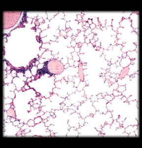
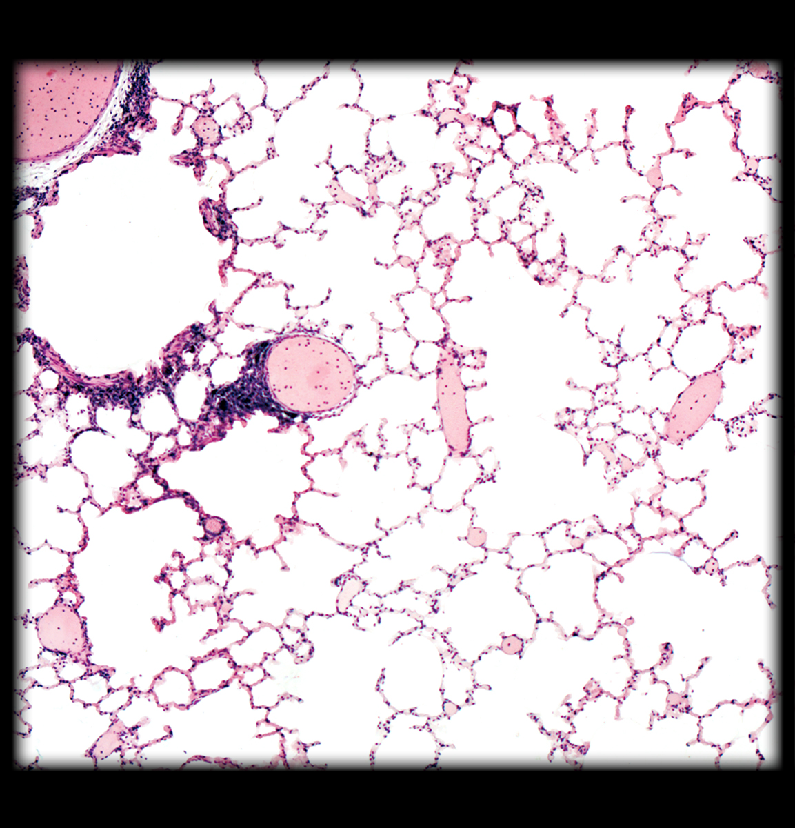
Exercise 36: Complete histology and human virtual respiratory dissection.
- Examine a microscopic section of lung tissue. See Figure 36.7 in your lab manual.
- Gross Anatomy: You are responsible for the being able to identify following respiratory structures from a cadaver photo or model on an exam. APR in Connect has a specific list of cadaver photos located in Anatomy & Physiology Revealed, My Course Content, List (Respiratory ZfjZD) and Module-Respiratory. Make sure to look at the My Dissection and My Histology.
External nares, nasal cavity, trachea, larynx, epiglottis, thyroid cartilage, hyoid bone (see skeletal module), primary (main) bronchi, pleural cavity, parietal pleura, visceral pleura, lungs, and diaphragm (muscular module).
DIGESTIVE ANATOMY:
Exercise 38: Complete histology and human digestion dissection.
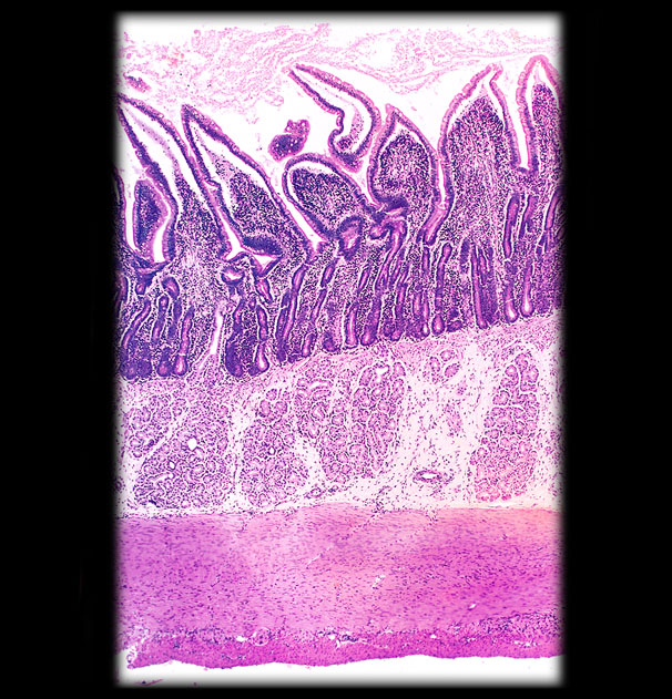
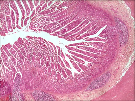
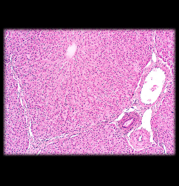
1. Observe microscopic sections shown below in this order: stomach, small intestine (duodenum and ileum shown), pancreas, liver and colon.
See Figures 38.6, 38.9, 38.16, 26.3, 27.3c in Marieb. 


- Gross Anatomy: You are responsible for the being able to identify following digestive structures from a cadaver photo or model on an exam. APR in Connect has a specific list of cadaver photos located in Anatomy & Physiology Revealed, My Course Content, List (Digestive tGZgk) and Module-Digestive. Make sure to look at the My Dissection and My Histology.
Parotid salivary gland and duct, teeth, oral cavity, hard palate, soft palate, tongue, pharynx, esophagus, peritoneal cavity, parietal peritoneum, visceral peritoneum,liver, greater omentum, gall bladder, stomach [cardia, fundus, body, pylorus], greater and lesser curvature of stomach, lesser omentum, pancreas, spleen (in lymphatic module), common bile duct, small intestine [duodenum, jejunum, ileum], mesentery proper, large intestine including: cecum, appendix, colon [ascending, transverse, descending, sigmoid], rectum, anus.
WEEK 10, OCTOBER 19-25 – ANATOMY OF URINARY AND REPRODUCTIVE SYSTEMS
Exercises 42 and 43: Complete histology and human virtual dissection.
1. Observe microscopic sections shown below of ovary and testis. Refer to Figures 42.2 and 43.6.
Ovary. Note the various levels of oocyte development within the follicles.
Testes. This is a cross section of the seminiferous tubules in the testes, which is the site of sperm formation. Note that the more immature spermatogonia and spermatocytes are towards the periphery of the tubules. The more mature cells are towards the center lumen of the tubule. In fact if you look closely, you can see the tails of the sperm in the lumen.
Gross Anatomy or Urinary and Reproductive Systems: You are responsible for the being able to identify following urinary and reproductive structures from a cadaver photo or model on an exam. APR in Connect has a specific list of cadaver photos located in Anatomy & Physiology Revealed, My Course Content, List (Urinary/Reproductive bKYQn and Modules-Urinary or Reproductive). Make sure to look at the My Dissection and My Histology.
Urinary: Kidney, renal capsule, renal cortex, renal medulla, renal pelvis, hilum (hilus), ureter, urinary bladder, and urethra.
Reproduction Male: Penis, testes, spermatic cord, ductus deferens (=vas deferens), prostate gland.
Reproduction Female: Uterus, uterine tube (=oviduct=fallopian tube), ovary, vagina, cervix, and vulva (containing vestibule and vaginal orifice).
WEEK 11, OCTOBER 26-NOVEMBER 1 – RESPIRATORY PHYSIOLOGY **NOTE LAB ASSIGNMENT IS DUE THIS WEEK**
Exercise 37: Complete the Virtual Lab called Pulmonary Function. It is found in the Set Three Laboratory Exercises and Exam heading. It’s completion is worth 20 points and is DUE November 1 at 11:59 pm. Lab exam 3 will have questions from this lab on it.
Note: Since you cannot go back into your Virtual Lab for studying reasons, the key points from the pulmonary virtual lab may be found here to help you prepare for Lab Exam Three: Virtual Lab Pulmonary Function
WEEK 12, NOVEMBER 2-8 – URINALYSIS
Exercise 41: Watch this clip from APR to see how urine is formed. http://anatomy.mheducation.com/html/apr.html?animal=human&id=4750
Then, watch this video on performing a urinalysis and then interpret the following 2 samples. Know what is normal and what is not for the following parameters for your lab exam. If a value is not normal, offer reasons why.
Sample One:
Color – clear and pale yellow
pH 6
Specific Gravity 1.005
Glucose and Ketones Negative
Protein Negative
Erythrocytes and Hemoglobin Negative
Leukocytes Negative
Sample Two:
Color red and cloudy
pH 5
Glucose and Ketones Positive
Specific Gravity 1.030
Protein weakly positive
Erythrocytes and Hemoglobin Positive
Leukocytes Negative
WEEK 13, NOVEMBER 9-15 – REPRODUCTIVE PHYSIOLOGY
Exercises 43 and 44: Watch the following videos. There will be lab exam questions from the videos.
WEEK 14, NOVEMBER 16-29 (at 11:59 pm) – EXAM 3
(25 questions, 50 points)
You will have 45 minutes in one sitting to complete this lab exam online in Connect. You will find it in the Set Three Laboratory Exercises & Exams Section. It consists of labeling and multiple choice questions. It only covers material after Lab Exam 2 (ie not comprehensive). Read the questions carefully, because a few have multiple answers. There is a 5% deduction in your score for each day you are late taking the exam after the due date.